ORIGINAL RESEARCH REPORTS
Plasticity of Calcium Signaling Cascades in Human Embryonic Stem Cell-Derived Neural Precursors
Oksana Forostyak,1Nataliya Romanyuk,2Alexei Verkhratsky,3Eva Sykova,2,4and Govindan Dayanithi 1,5
Human embryonic stem cell-derived neural precursors (hESC NPs)are considered to be a promising tool for cell-based therapy in central nervous system injuries and neurodegenerative diseases.The Ca 2+ion is an important intracellular messenger essential for the regulation of various cellular functions.We investigated the role and physiology of Ca 2+signaling to characterize the functional properties of CCTL14hESC NPs during long-term maintenance in culture (in vitro).We analyzed changes in cytoplasmic Ca 2+concentration ([Ca 2+]i )evoked by high K +,adenosine-5¢-triphosphate (ATP),glutamate,g -aminobutyric acid (GABA),and caffeine in correlation with the expression of various neuronal markers in different passages (P6through P10)during the course of hESC differentiation.We found that only differentiated NPs from P7exhibited signi?cant and speci?c [Ca 2+]i responses to various stimuli.About 31%of neuronal-like P7NPs exhibited spontaneous [Ca 2+]i oscillations.Pharmacological and immunocytochemical assays revealed that P7NPs express L-and P/Q-type Ca 2+channels,P2X 2,P2X 3,P2X 7,and P2Y purinoreceptors,glutamate receptors,and ryanodine (RyR1and RyR3)receptors.The ATP-and glutamate-induced [Ca 2+]i responses were concentration-dependent.Higher glutamate concentrations (over 100m M)caused cell death.Responses to ATP were observed in the presence or in the absence of extra-cellular Ca 2+.These results emphasize the notion that with time in culture,these cells attain a transient period of operative Ca 2+signaling that is predictive of their ability to act as stem elements.
Introduction
H
uman embryonic stem cells (hESCs)are pluripotent cells derived from the inner cell mass of a preimplanta-tion embryo [1].In vitro,these cells are able to maintain a normal euploid kariotype,differentiate into derivatives of all 3germ layers,and proliferate extensively [2,3].These prop-erties make them unique candidates for cell transplantation,research into growth factors and early human development,and for drug discovery.Substantial progress has been made recently in the differentiation of hESCs into a neuronal phe-notype [3–5],this being a promising strategy for cell-based therapy of central nervous system injuries and neurodegen-erative diseases.However,to date,several important ques-tions remain unanswered:(1)when and at what stage of differentiation should cells be transplanted;(2)what are the functional properties (ion channels,receptors,and second messengers)of these cells and how are they regulated;and (3)how compatible are these properties with the physiological or pathological environment at the site of transplantation and
treatment?Hitherto,with a few exceptions,the quality of stem cells is generally evaluated by determining the expression of various genes and key proteins during the process of differ-entiation;these however,although being present in the cell,may be physiologically inactive.Therefore,the aim of this study was,for the ?rst time,to determine and characterize Ca 2+signals activated by physiological stimulation of neural precursors (NPs)derived from hESCs.
Ca 2+is a ubiquitous second messenger involved in the regulation of almost all known cellular processes and,above all,in de?ning the life and death of every cell [6–10].Signals mediated by Ca 2+are fundamental for fertilization,cell differentiation,proliferation [11],intercellular signaling,transcription factor activation,and various death programs including necrosis and apoptosis [12].Ca 2+can enter the cytoplasm from 2sources:either by an in?ux via plasma-lemmal voltage-operated and receptor-operated Ca 2+chan-nels (VOCC and ROCC respectively)or by release from intracellular stores,such as the endoplasmic reticu-lum,through endomembrane Ca 2+channels classi?ed as
Departments of 1Molecular Neurophysiology,and 2Neuroscience,Institute of Experimental Medicine,Academy of Sciences of the Czech Republic,Prague,Czech Republic.3
School of Biological Sciences,University of Manchester,Manchester,United Kingdom.4
Department of Neuroscience,Second Medical Faculty,Charles University,Prague,Czech Republic.5
Institut National de la Sante
′et de la Recherche Me ′dicale,Unite ′de recherche U710,Universite ′Montpellier 2,Montpellier;and Ecole Pratique des Hautes Etudes,Paris,France.
STEM CELLS AND DEVELOPMENT Volume 22,Number 10,2013óMary Ann Liebert,Inc.DOI:10.1089/scd.2012.0624
1506
inositol-1,4,5-trisphosphate receptors(InsP3Rs)and ryano-dine receptors(RyRs).The variety of functions executed by Ca2+depends on the speed,amplitude,and spatiotemporal pattern of Ca2+signals and by interactions between Ca2+ and other signaling pathways[9].For example,changes in [Ca2+]i following the activation of purinoceptors(P2X3, P2X4,P2Y1,and P2Y2)promote cell proliferation in murine ESCs[13].On the other hand,glial excitability depends on Ca2+waves that often occur as a result of adenosine-5¢-tri-phosphate(ATP)-mediated signaling through P2Y receptors [14].The entry of Ca2+through VOCC and the release of Ca2+from internal stores modulate neuronal excitability [15].A transient increase in[Ca2+]regulates cellular secre-tion and cellular motility during neuronal development[16]. Both g-aminobutyric acid(GABA)and glutamate have been shown to in?uence NP cell proliferation during development [17,18].Hence,a detailed characterization of the Ca2+sig-naling cascades that are activated by various stimuli is useful in determining the functional state of the cell and may even predict its fate.
In the previous work we have described a novel protocol for the ef?cient generation of NPs from hESCs[19].Our re-sults showed that(1)hESC NPs are able to differentiate into a neuronal phenotype and to develop into functionally active neurons;(2)P8hESC NPs are the appropriate candidate for transplantation compared to undifferentiated hESCs and to P1,P5,and P10;(3)long-term maintenance in vitro decreases tumorogenisity,although simultaneously attenuates prolif-erative activity and differentiation potential.Our study fur-ther revealed that the pro?le of NPs changes with the length of maintenance in culture.
To date,the role of Ca2+,its homeostasis and signaling potential in ESCs differentiated to a neuronal phenotype have not been studied.To our knowledge there are only a few reports available on mouse ESCs[20,21]and on hESCs differentiated toward neurones[22–25].Therefore,we un-dertook the present study to determine whether the func-tional properties of NPs change during long-term maintenance in culture.We also asked at what stage of dif-ferentiation are these cells in ideal physiological condition? We identi?ed and analyzed molecular cascades of[Ca2+]i homeostasis and Ca2+signaling in correlation with hESC differentiation into a neuronal phenotype.
Materials and Methods
Drugs and solutions
Chemicals obtained from Sigma-Aldrich:accutase,lami-nin,cadmium chloride,nickel chloride,nicardipine hydro-chloride,ATP,a,b-Methyleneadenosine5¢-triphosphate lithium salt(a,b-MeATP),suramin sodium salt,pyridoxal phosphate-6-azo(benzene-2,4-disulfonic acid)tetrasodium salt hydrate(PPADS),2¢(3¢)-O-(4-Benzoylbenzoyl)adenosine 5¢-triphosphate triethylammonium salt(BzATP),l-Glutamic acid potassium salt monohydrate,N-Methyl-D-aspartic acid (NMDA),kainic acid,GABA,adenosine,b-mercaptoethanol, bovine serum albumin,and Triton-X100;from GIBCO: Dulbecco’s modi?ed Eagle’s medium(DMEM)/F12,l-glutamine,penicillin/streptomycin,fetal bovine serum, collagenase type IV,and B27;from Invitrogen:human re-combinant?broblast growth factor(hrFGF),Fura-2AM 1mM solution in anhydrous dimethyl sulfoxide(DMSO) cell permeant,and4¢,6-diamidino-2-phenylindole dihydro-chloride(DAPI);from R&D Systems:human recombinant epidermal growth factor(hrEGF);from Molecular Probes (Eugene,OR):goat anti-mouse IgG conjugated with Alexa-Fluor594,goat anti-rabbit IgG conjugated with Alexa-Fluor 488,and Pluronic F-127;from Tocris Bioscience:NF279; from Polysciences,Inc.(Warrington,PA):Aqua Poly/Mount mounting medium;from Alomone Labs Ltd.:caffeine,rya-nodine,cyclopiazonic acid(CPA),o-conotoxin MVIIC (MVIIC),and o-conotoxin GVIA(GVIA).
Concentrated stock solutions of nicardipine,glutamate, and ryanodine were prepared in DMSO,while the remaining stock solutions of agonists/antagonists were dissolved in dH2O.For each experiment,caffeine was freshly dissolved in Normal Locke’s buffer(NL)at37°C and vortexed until the caffeine crystals dissolved.All concentrated stock solutions were stored at-20°C.Test solutions were daily prepared using aliquots from frozen stocks to obtain the working concentrations.All buffers and solutions in this study were made using ion-free dH2O from Merck,Germany.
hESC and hESC NP culture
hESCs line CCTL14(complete information on the deriva-tion and characterization of CCTL14line of hESCs is avail-able at https://www.doczj.com/doc/1a12412278.html,/science/sclines.htm)were cultured on a feeder layer of mitomycin-C-treated mouse embryonic ?broblasts in gelatin-coated tissue culture dishes.The culture medium(CM)was(D-MEM/F-12without l-glutamine) supplemented with15%fetal bovine serum,1%nonessential amino acids,2mM l-glutamine,penicillin,and streptomycin at50U/mL,0.1mM b-mercaptoethanol,and4ng/mL hrFGF.Colonies of hESCs were passaged every4–7days using either mechanical scraping with a glass pipette to provide low-density cultures of undifferentiated cells or en-zymatic dissociation with collagenase type IV to provide high-density cultures of undifferentiated cells[19].
NPs were derived from the CCTL14line of human ESCs as described previously[19].Brie?y,to induce neural differen-tiation,clumps of undifferentiated hESCs were plated in agarose-coated tissue culture dishes with CM without hrFGF for4days,then with CM supplemented with Noggin for the next4days.At this time,70%–90%of the colonies formed embryoid bodies(EBs).Aggregates whose diameter ex-ceeded0.5mm were dissected into smaller clumps with20G surgical blades,then replaced in serum-free medium or NP medium(NPM)and cultured for6days.At this stage,cells in the EBs were de?ned as passage 1.NPM consisted of DMEM/F12medium(1:1),B27supplement(1:50),2mM l-glutamine and penicillin,and streptomycin at50U/mL, supplemented with20ng/mL hrEGF and20ng/mL hrFGF. For the long-term propagation of hESC NPs,the EBs were dissociated by accutase and the cells were plated onto laminin-coated culture dishes.NPs were cultured in NPM and passaged by accutase each5–7days.
Antibodies and immunocytochemistry
Cells plated onto laminin-coated coverslips were washed in phosphate-buffered saline(0.1M PBS,pH7.2)and?xed with4%paraformaldehyde in PBS for15min.The?xed cells
CALCIUM SIGNALING IN HUMAN EMBRYONIC STEM CELLS1507
were washed twice in PBS prior to immunostaining.Per-meabilization and blocking were carried out in a blocking buffer consisting of 0.4%Triton-X 100and 10%bovine serum albumin in 0.1M PBS for 45min at 22°C.Primary antibodies (See Table 1)were diluted in buffer consisting of 0.1%Triton-X 100and 2%bovine serum albumin in PBS overnight at 4°C.After 2washes with PBS,appropriate secondary antibodies (Alexa Fluor 488-conjugated goat anti-rabbit IgG [1:200]or Alexa Fluor 594-conjugated goat anti-mouse IgG [1:200]),were applied for 30min at 22°C.To visualize the cell nuclei,the coverslips were incubated with 300nM DAPI in PBS for 5min at 22°C.Finally,the coverslips with cells were mounted using Aqua Poly/Mount mounting medium and examined using a ZEISS LSM 510DUO confocal microscope (Carl Zeiss).
Dye loading and measurements of intracellular [Ca 2+]i
[Ca 2+]i measurements on single cells were performed as previously reported [26–31].Brie?y,cultures of hESC NPs,plated on 24mm glass-bottom dishes (WillCo-dish,WillCo Wells B.V,Amsterdam,Netherlands)coated with laminin,were incubated with 2.5m M Fura-2AM with 0.02%Pluronic F-127in CM at 37°C and 5%CO 2for 40min.Loaded cells were then washed and the CM replaced with NL buffer containing (in mM):NaCl,140;KCl,5;MgCl2,1.2;CaCl 2,2.2;glucose,10;and HEPES-Tris,10;pH 7.25and kept at 37°C throughout the time course of the experiment.The osmolarity of the buffer was 298–300mosmol/l -1.Fluorescence mea-
surements of [Ca 2+]i were performed with a fast ?uorescence microspectro?uorimetry system based on an inverted micro-scope (Axiovert,Zeiss)equipped for epi?uorescence (Plan-Neo?uar 100x/1.30oil immersion objective).To achieve fast switching between different excitation wavelengths,a rotating ?lter wheel was mounted in the excitation light path.The cells were alternately illuminated (200Hz)at 340–10nm and 380–10nm.To minimize the background noise of the Fura-2signal,successive values were averaged to a ?nal time reso-lution of 320ms.A measuring ampli?er was synchronized to the ?lter wheel to measure the ?uorescence intensities re-sulting from different wavelengths.The FFP software con-trolled the acquisition of the intensity data and provided functions for adjusting the signal values and the display and storage of the measured data.A CCD camera was used to visualize the cells.The [Ca 2+]i measurement values are ex-pressed as the ratio units (RU)between the ?uorescence ob-tained with 2excitation wavelengths,340nm (A)and 380nm (B).Fura-2calibration was performed in these cells in vitro following the procedure described by Lambert et al.[32]and Jamen et al.[33],which yielded R min =0.08,R max =2.02,b =1.757,and K d =224at 37°C.
Drug application
As described previously [28,34,35],the control and test solutions were applied using a temperature controlled multi-channel polypropylene capillary perfusion system (Warner Instruments,Inc.).A single outlet capillary tubing (100m m
Table 1.Antibodies Used for Immunocytochemistry
Antibody
Dilution Host/Antibody type Company
Secondary antibody used
Anti-NeuN
1:100Mouse monoclonal Merck Millipore Alexa Fluor 594goat anti-mouse IgG Anti-b -tubulin isotype III 1:1000Mouse monoclonal Sigma-Aldrich Alexa Fluor 594goat anti-mouse IgG Anti-MAP-2
1:1000Mouse monoclonal Merck Millipore
Chemicon
Alexa Fluor 594goat anti-mouse IgG Anti-nestin 1:2000Mouse monoclonal Merck Millipore Alexa Fluor 594goat anti-mouse IgG Anti-myelin/
oligodendrocyte speci?c protein 1:250Mouse monoclonal Merck Millipore
Chemicon Alexa Fluor 594goat anti-mouse IgG Anti-S1001:400Rabbit polyclonal DAKO Denmark Alexa Fluor 488goat anti-rabbit IgG Anti-GFAP
1:80Rabbit
Sigma-Aldrich Alexa Fluor 488goat anti-rabbit IgG Anti-glutamine synthetase 1:1000Mouse monoclonal Merck Millipore Alexa Fluor 594goat anti-mouse IgG Anti-glutamate 1:1000Mouse monoclonal Sigma-Aldrich Alexa Fluor 594goat anti-mouse IgG Anti-GABA 1:500Mouse monoclonal Sigma-Aldrich
Alexa Fluor 594goat anti-mouse IgG Anti-Iba11:1000Rabbit polyclonal Wako Chemicals GmbH Alexa Fluor 488goat anti-rabbit IgG Anti-NG2
1:400Rabbit polyclonal Merck Millipore
Alexa Fluor 488goat anti-rabbit IgG Anti-L-type Ca 2+CP a 1C 1:100Rabbit polyclonal Santa Cruz Biotechnology Alexa Fluor 488goat anti-rabbit IgG Anti-N type Ca 2+CP a 1B 1:100Rabbit polyclonal Santa Cruz Biotechnology Alexa Fluor 488goat anti-rabbit IgG Anti-P/Q-type Ca 2+CP a 1A 1:100Rabbit polyclonal Santa Cruz Biotechnology Alexa Fluor 488goat anti-rabbit IgG Anti-P2X21:100Rabbit polyclonal Alomone Labs Alexa Fluor 488goat anti-rabbit IgG Anti-P2X31:100Rabbit polyclonal Alomone Labs Alexa Fluor 488goat anti-rabbit IgG Anti-P2X41:100Rabbit polyclonal Alomone Labs Alexa Fluor 488goat anti-rabbit IgG Anti-P2X61:100Rabbit polyclonal Alomone Labs Alexa Fluor 488goat anti-rabbit IgG Anti-P2X71:100Rabbit polyclonal Alomone Labs Alexa Fluor 488goat anti-rabbit IgG Anti-RyR 31:100Rabbit polyclonal Alomone Labs Alexa Fluor 488goat anti-rabbit IgG Anti-RyR 21:200Rabbit polyclonal Alomone Labs Alexa Fluor 488goat anti-rabbit IgG Anti-RyR 11:200Rabbit polyclonal Alomone Labs Alexa Fluor 488goat anti-rabbit IgG Anti-NMDA e 4
1:100
Rabbit
polyclonal
Santa Cruz
Biotechnology
Alexa
Fluor
594
goat
anti-rabbit
IgG
GFAP,glial ?brillary acidic protein;GABA,g -aminobutyric acid;RyR,ryanodine receptor;NMDA,N-Methyl-D-aspartic acid.
1508
FOROSTYAK ET AL.
inner diameter)with a?ow rate of250m L/min was posi-tioned close to the tested cell(0.5mm).The selected cell was subjected to a constant?ow of control buffer or test solutions. Each capillary was fed by a reservoir45cm above the bath and connected to a temperature control device(Harvard).The temperature of all solutions was maintained at37°C.In this approach,switching the?ow from one capillary to the next resulted in complete solution exchange within1–3s.After each application of the tested drug,the cells were washed with control buffer.This method allowed for the fast and re-liable exchange of the solution surrounding the selected cell under observation without exposing the neighboring cells. Data analysis and statistical methods
Origin8.5.1was employed for plotting and statistical pro-cedures.The results are expressed as mean–SEM.The num-ber of the sample size(n)given is the number of cells tested according to the same protocol(control,test drug,and re-covery)for each group.The?gures(traces)show on-line single cell measurements of the[Ca2+]i levels before and after the application of test substances,while bar diagrams and numerical data are given as mean–S.E.M.and present the peak amplitude of the[Ca2+]i increase as a ratio between the ?uorescence values of340/380nm excitation wavelengths. The results were analyzed using one-way ANOVA.Differ-ences were considered statistically signi?cant if P£0.05. Results
[Ca2+]i responses in undifferentiated hESC Undifferentiated hESCs(n=34)from2independent cul-tures were subjected to the application of K+(50mM),ATP (100m M),glutamate(50m M),GABA(10m M),and CPA (10m M).These cells were only partially responsive to ATP. To identify the speci?c involvement of the type(s)of active purinergic receptors in undifferentiated hESC,we tested the effect of adenosine diphosphate(ADP;100m M)and adeno-sine(2m M).ADP had no effect,whereas adenosine caused rise in[Ca2+]i in only18%(n=11).P2receptor antagonist suramin(300m M)inhibited ATP-induced[Ca2+]i by 33.3%–23%(n=4),PPADS(10m M)had no effect(n=3). [Ca2+]i responses in hESC NPs change during
long term propagation in culture
The functional properties of hESC NPs were studied during long-term propagation in vitro starting from P6NPs to P10NPs.All data within each passage were collected and analyzed from a minimum of3independent experiments. Cells were chosen randomly and subjected to the same ex-perimental protocol.The cells from each passage were sub-jected to various stimuli such as K+(50mM),ATP(100m M), glutamate(50m M),GABA(10m M),and caffeine(20mM; Table2).The results indicate that P6NPs were sensitive to the application of glutamate and K+(27%–1.3%and 11%–1%,respectively).Only a few cells were sensitive to other stimuli such as GABA or ATP(7%–1%each)and to caffeine(4%–2%).The percentages of P7cells responding to K+,ATP,glutamate,GABA,and caffeine were50%–3.9%, 46%–2.2%,57%–0.3%,5%–2.3%,and18%–2.1%,respec-tively.Similarly,the percentages of P8NPs responding to K+,ATP,glutamate,GABA,and caffeine were32%–4%, 24%–3.5%,9%–1.3%,16%–1.1%,and16%–1%,respec-tively.The cells from P9were responsive to ATP(9%–1.9%) and nonresponsive to GABA.Less than5%of cells were sensitive to other stimuli.P10NPs were not sensitive to glutamate or GABA,while5%–2.3%of cells were sensitive to ATP and2.3%(1out of44cells tested)to K+or caffeine. Immunocytochemical staining for Ca2+channels and RyRs revealed the expression of L-type and P/Q-type VOCC, ryanodine receptors RyR1and RyR3,and the weak expres-sion of RyR2in all passages(Table3).Since the number of cells responding to physiological stimuli was signi?cantly higher in passage P7NPs(59%–2.5%)than in other pas-sages(P6NPs:41%–1.6%;P8NPs:49%–1.1%;P9NPs: 15%–1.7%;and P10NPs:7%–0.2%),these cells were chosen for further experimentation in this study. Characterization of[Ca2+]i transients mediated
by Ca2+channels in P7hESC NPs
We monitored Ca2+entry through VOCC as[Ca2+]i transients evoked by depolarization with50mM K+.The application of high K+solution evoked a rapid increase in [Ca2+]i in50%(n=145)of the tested cells.Preincubation with Cd2+(100m M),a nonspeci?c blocker of high-voltage acti-vated(HVA)Ca2+channels(L-N-P-Q-and R-types),to-gether with Ni2+(50m M),a more speci?c blocker of low-voltage activated Ca2+channels(T-type),for5min signi?-cantly reduced the[Ca2+]i responses induced by K+in all cells tested by85.3%–7.5%(n=7)indicating the involvement of voltage-activated Ca2+channels in depolarization-induced Ca2+entry(Fig.1A,B).To further characterize the involvement of speci?c subtypes of HVA Ca2+channels,we used speci?c Ca2+channel blockers such as nicardipine(for
Table2.The Number of Cells Responding to Various Physiological Stimuli in Different Passages of Human Embryonic Stem Cell-Derived Neural Precursors(P6–P10NP s)
P6NPs P7NPs P8NPs P9NPs P10NPs Number of cells tested n=44n=44n=45n=45n=44 Cells sensitive to1or more of applied stimuli41%–1.6%59%–2.5%49%–1.1%15%–1.7%7%–0.2% K+50mM11%–1%50%–3.9%32%–4%4%–2.2% 2.3% ATP100m M7%–1%46%–2.2%24%–3.5%9%–1.9%5%–2.3% Glutamate50m M27%–1.3%57%–0.3%9%–1.3%4%–2.2%0 GABA10m M7%–1%5%–2.3%16%–1.1%00 Caffeine20mM4%–2%18%–2.1%16%–1.1%4%–2.1% 2.3% ATP,adenosine-5¢-triphosphate.
CALCIUM SIGNALING IN HUMAN EMBRYONIC STEM CELLS1509
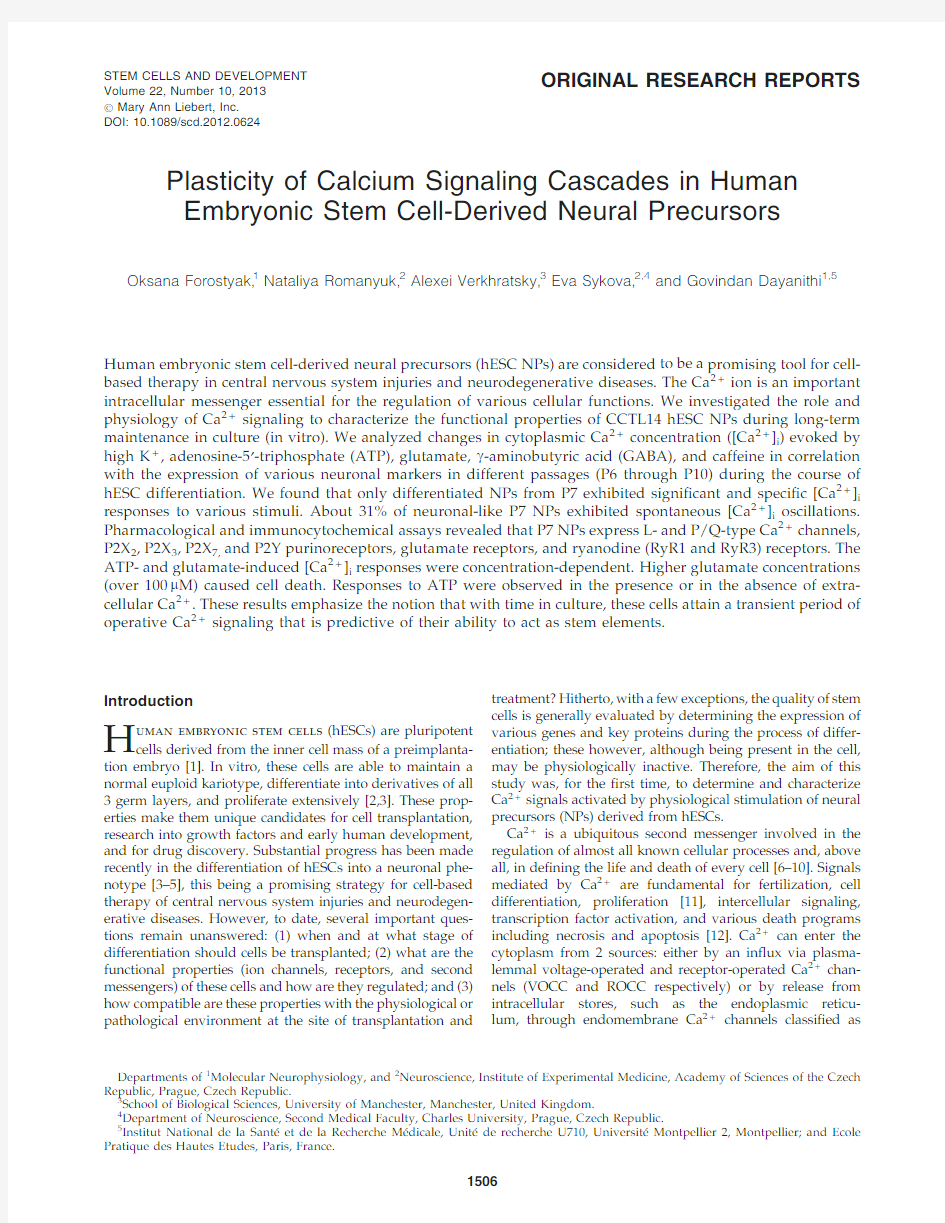
L-type),MVIIC(for P/Q type),and GVIA(for N-type). Application of10m M nicardipine reduced[Ca2+]i responses in67%of tested P7NPs(n=12).In2out of8cells nicardipine totally abolished the[Ca2+]i responses(Fig.1C,D).In the other6cells,it signi?cantly reduced the amplitude of the responses by54%–19.5%(P=0.03;n=6),suggesting the contribution of L-type Ca2+channels in P7NPs.The appli-cation of300nM MVIIC(Fig.1E,F),which is known to block the P/Q-type of VOCC[36],reduced the[Ca2+]i responses by92%–32%(P=0.004;n=5)in all cells.The application of MVIIC at a higher concentration(1m M),which was reported to also block N-type Ca2+channels,completely inhibited the K+-induced responses in the tested cells(n=3).Further,we used another speci?c N-type VOCC blocker,GVIA,at2 different concentrations(500nM and800nM),which only partially and reversibly reduced the K+-induced responses by20%–14.6%(P=0.47;n=11)and48%–31.8%(P=0.27; n=9),respectively.To con?rm the above[Ca2+]i measure-ment results,we performed a series of immunocytochemical analyses.No positive immunostaining for the a1B subunit of the N-type of VOCC was observed,but positive im-munostaining for the a1C subunit of the L-type of VOCC (Fig.1G)and the a1A subunit of the P/Q-type of VOCC(Fig. 1H)was observed,suggesting the presence of L-and P/Q-type Ca2+channels in P7NPs.
[Ca2+]i signaling through purinergic
receptors in P7hESC NPs
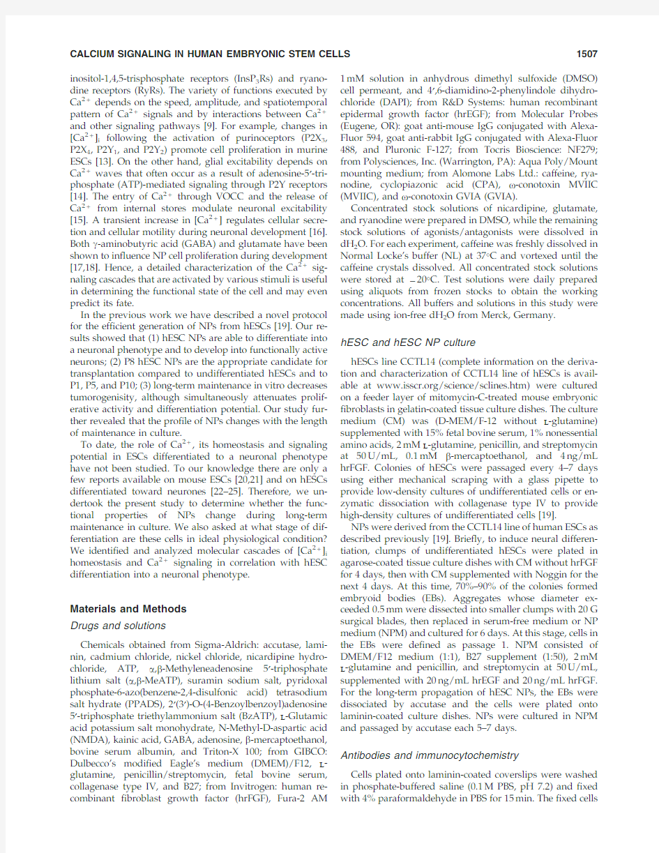
The application of ATP(100m M)induced a rapid[Ca2+]i increase in33out of71(47%)NPs tested with the mean amplitude of0.8–0.12(RU)(Fig.2A).The rise in[Ca2+]i in response to ATP was signi?cantly inhibited(by65%–25%)by the broad-spectrum P2receptor antagonist suramin(300m M) (P=0.01;n=8)(Fig.2B,C).Another P2receptor antagonist, PPADS,had a similar effect at a concentration of10m M,de-creasing the[Ca2+]i response to ATP by73%–20%(P=0.02; n=8),(Fig.2D,E).These results suggest that functional P2 purinoceptors are present in P7NPs.
To further identify which P2receptors(ionotropic P2X or metabotropic P2Y)operate in hESC NPs,we studied the ef-fect of the P2X agonist a,b-meATP.In64%of the cells,the application of a,b-meATP(100m M)transiently increased [Ca2+]i,by a mean amplitude of0.86–0.14,n=16(Fig.2A). The application of20m M BzATP,another P2X receptor ag-onist,evoked a[Ca2+]i increase in18%of the tested cells with the mean amplitude of0.28–0.07,(n=22;Fig.2A).The P2X receptors antagonist,NF279,was ineffective at concen-trations from20nM to1m M at which it is speci?c agonist for P2X1receptors(Fig.2F),whereas at a higher concentration (100m M),it inhibited the ATP-induced[Ca2+]i increase by 69%–20.6%,P=0.04;n=3(Fig.2G).Immunocytochemical staining with antibodies directed against P2X2,P2X3,P2X4, P2X6,and P2X7receptors revealed expression of P2X2(Fig. 2H),P2X3(Fig.2I)and P2X7R(Fig.2J).Finally,to investigate P2Y-mediated Ca2+signaling,the effects of ATP were ex-amined in Ca2+-free medium.In the absence of extracellular Ca2+,the application of ATP induced an increase in[Ca2+]i in86%of the tested cells with a mean amplitude of 0.34–0.15,n=7(Fig.2A).The application of100m M ADP,a P2Y1and P2Y12agonist,elevated[Ca2+]i by0.38–0.08(n=5) in56%of the tested cells(Fig.2A).These data suggest the involvement of both the P2X(P2X2,P2X3,and P2X7)and P2Y purinoceptors in Ca2+signaling in P7NPs.
Ca2+release from intracellular Ca2+stores and spontaneous[Ca2+]i transients in P7hESC NPs
To check for functional intracellular Ca2+stores in P7NPs, we used caffeine(20mM),which in millimolar concentra-tions acts as an inhibitor of intracellular receptors for IP3 while being a potent activator of RyR.We also applied rya-nodine at1m M,the concentration that opens the RyR.A brief application(10s)of either caffeine or ryanodine induced a [Ca2+]i rise in18%(n=44)and22%(n=9)of P7hESC NPs, respectively.Immunostainings for RyR1(Fig.3A)and RyR3 (Fig.3B)were positive in all tested cells.The application of 10m M CPA,a potent,selective,and reversible inhibitor of the sarco-endoplasmic reticulum Ca2+-ATP ase pump,caused an intracellular[Ca2+]i increase in12out of13tested cells(92%) with a mean amplitude of0.38–0.05.
Spontaneous[Ca2+]i transients were observed in51out 164of tested cells(31%)in the absence of any stimuli(Fig. 3C).The amplitude of the spontaneous[Ca2+]i transients was0.74–0.05(n=91),and they appeared with a mean fre-quency of1per3.2min.These transients were partially in-hibited by the application of VOCC blockers(100m M Cd2+, 50m M Ni2+,and10m M nicardipine)or completely by the removal of extracellular Ca2+(Fig.3D).
[Ca2+]i responses to glutamate in P7hESC NPs
We tested the[Ca2+]i responses of P7NPs to various concentrations of glutamate(1m M–1mM;n=118).Out of 118,the glutamate caused an elevation of[Ca2+]i in80cells
Table3.Expression of Various Neural Markers and Ca2+Sensitive Channels in P6–P10hESC NPs
P6 NPs
P7
NPs
P8
NPs
P9
NPs
P10
NPs
Neural markers
Nestin++++++++++
NeuN++++++++
b III tubulin+++++++++++++
MAP2-----
Glutamate++++---
GABA+-+--
GFAP++++++++-
S100+++++++++
OLIG-++---
GS-++---
NG2----+
Ca2+-sensitive channels
L-type VOCC+++++++++++
N-type VOCC-----
P/Q-type VOCC+++++++++++
RyR1+++++++++++++++
RyR2+++++
RyR3+++++++++++
‘‘-’’-negative immunostaining.
‘‘+’’-faint positive immunostaining/few cells express the marker.
‘‘++’’-positive immunostaining/many cells express the marker.
‘‘+++’’-very positive/majority of cells express the marker.
VOCC,voltage-operated Ca2+channels.
1510FOROSTYAK ET AL.
FIG. 1.Voltage-operated Ca 2+channels in P7human embryonic stem cell-derived neural precursors (hESC NPs).Example of traces (A,C,E)from individual cells for each experimental design show the block of K +responses by speci?c Ca 2+channel antagonists.The cells were ?rst exposed to control K +depo-larization and the [Ca 2+]i responses were monitored.Note that the am-plitude of the control responses to high K +is identical without any run down.Subsequently,the same cell was preincubated for 5min with Ca 2+channel blockers [Cd 2+/Ni 2+;Nic:nicardipine and o -con-otoxin MVIIC (MVIIC)]as indi-cated on the trace and then again challenged with high K +.(B)Pre-incubation of cells with 100m M Cd 2+together with 50m M Ni 2+signi?cantly reduced [Ca 2+]i re-sponses in all tested cells (n =7).The trace (C)and bar diagram (D)show the reduction of the high K +-induced [Ca 2+]i responses by the L-type Ca 2+channel blocker 10m M nicardipine (n =6).The trace (E)shows the K +-induced [Ca 2+]i re-sponses in the presence of and after preincubation with the P/Q-type Ca 2+channel blocker,MVIIC (300nM).(F)Bar diagram shows the MVIIC inhibition of the high-K +responses (n =5).G,H are confocal images of P7hESC NPs,co-stained for b III tubulin (G,H)(middle panel )and for L-type Ca 2+CP a 1C (H-280)(G),(left panel )or P/Q-type Ca 2+CP a 1A (H-90)(H),(left panel ).Merged images are pre-sented in the right panels (G,H).Scale bars =20m m.*P =0.05;**P =0.005;***P =0.0005.Color images available online at www.liebertpub .com/scd
CALCIUM SIGNALING IN HUMAN EMBRYONIC STEM CELLS 1511
FIG.2.Purinergic responses in P7hESC NPs.P7NPs express functional P2X and P2Y purinergic receptors.(A)Bar diagram showing the mean amplitude of the [Ca 2+]i increase in response to various purinergic receptor agonists in P7hESC NPs.The cells were exposed to100m M adenosine-5¢-triphosphate (ATP),100m M a ,b -Methyleneadenosine 5¢-triphosphate lithium salt (a ,b -meATP),20m M 2¢(3¢)-O-(4-Benzoylbenzoyl)adenosine 5¢-triphos-phate triethylammonium salt (BzATP),and 100m M adenosine diphosphate (ADP).The application of ATP also re-sulted in a rise in [Ca 2+]i in the absence of external Ca 2+.(B,D)Representative tra-ces show the ATP-induced [Ca 2+]i re-sponses obtained in the presence of and after the washout of the purinergic recep-tor antagonists suramin and pyridoxal phosphate-6-azo(benzene-2,4-disulfonic acid)tetrasodium salt hydrate (PPADS).Incubation with 300m M suramin or 10m M PPADS after control ATP application sig-ni?cantly inhibited the ATP responses.(C,E)Bar diagrams showing the amplitude of the [Ca 2+]i response to ATP before (con-trol)and after incubation with the antag-onists (C)300m M suramin (*P =0.01,n =8)and (E)10m M PPADS (*P =0.02,n =8).(F,G )Bar diagrams showing the effect of NF279in P7NPs.At 1m M concentration NF279had no effect (F),while at 100m M concentration (G)it signi?cantly inhibited ATP-induced [Ca 2+]i responses (*P =0.04;n =3).Confocal images (H,I,J)showing the staining for P2X 2(H),P2X 3(I),and P2X 7(J)receptors.Cell nuclei are visual-ized with 4¢,6-diamidino-2-phenylindole dihydrochloride (DAPI)staining.Scale bars =20m m.Color images available on-line at https://www.doczj.com/doc/1a12412278.html,/scd
1512FOROSTYAK ET AL.
(68%).Glutamate at 1mM caused cell death,which was determined by the loss of Ca 2+signals (Supplementary Fig.S1;Supplementary Data are available online at www https://www.doczj.com/doc/1a12412278.html,/scd).Therefore,the concentration-dependent elevation of [Ca 2+]i was determined for the doses ranging
between 1and 100m M.The amplitude of the glutamate-induced [Ca 2+]i responses at various concentrations ranged,respectively at 1m M =0.27–0.04,n =6;at 10m M =0.29–0.04,n =6;at 50m M =0.56–0.15,n =29;and at 100m M =0.95–0.06,n =31(Fig.4A,B).When the cells were exposed brie?y,10
s,
FIG.3.Ca 2+release from intracellular Ca 2+stores and spontaneous [Ca 2+]i transients in P7hESC NPs.(A,B)Confocal images of P7hESC NPs.(A)Ryanodine receptor (RyR)1and (B)RyR 3receptors are present in all cells and are co-localized with immunostaining for b III tubulin.Scale bars =20m m.(C)A representative trace showing spontaneous [Ca 2+]i oscillations observed in the same cell.These types of oscillations were observed in about 31%of tested P7hESCs.(D)Traces showing the inhibition of spontaneous [Ca 2+]i activity after removal of extracellular Ca 2+,application of voltage-operated Ca 2+channels (VOCC)blockers Cd 2+(100m M)and Ni 2+(50m M)and L-type VOCC blocker nicardipine (10m M).Color images available online at https://www.doczj.com/doc/1a12412278.html,/scd
CALCIUM SIGNALING IN HUMAN EMBRYONIC STEM CELLS 1513
to 1mM glutamate,there was a robust increase in [Ca 2+]i ,the Ca 2+level was high,sustained without any decay,and did not return to resting level even after wash of glutamate for a longer duration (Supplementary Fig.S1.)This phe-nomenon was observed in all 8cells tested.Glutamatergic signals in the nervous system are mediated by ionotropic [NMDA,a -amino-3-hydroxy-5-methyl-4-isoxazolepropionic acid (AMPA),and kainate]and metabotropic (mGlu)recep-tors.Two out of 9tested cells were sensitive to the applica-tion of NMDA (100m M),while only 1out of 6cells was sensitive to kainic acid (100m M).To determine the contri-bution of metabotropic glutamate receptors,we applied glutamate in the absence of extracellular Ca 2+;this caused a [Ca 2+]i increase in only 1out of 9cells suggesting that the sensitivity of P7NPs to glutamate is mediated mainly by ionotropic glutamate receptors.
Expression of neural markers in NPs derived from hESC
The expression of various neural markers was analyzed in passages 6–10(P6–P10)NPs.Table 1shows the list of pri-mary and secondary antibodies used.All immunocyto-chemical results were obtained from 2independent stainings with each antibody.The results from P6–P10are summa-rized in Table 3.The expression of various neural markers in P7NPs is shown in Fig.5.The neuronal markers NeuN (Fig.5Ba)and b III tubulin (Fig.5Ca,Da,Ea,Fa)were expressed in all passages,with the highest expression in P6–P8.Im-munostaining for neuronal marker MAP-2was negative in all passages.Staining for nestin (Fig.5Aa),a marker of neural progenitor cells,revealed the presence of numerous nestin-positive cells in all passages of hESC.Glial cells markers such as glial ?brillary acidic protein (GFAP)(Fig.5Db,Gb)and astrocytic marker S100(Fig.5Ab,Bb,Cb,Hb)were also expressed throughout all passages except GFAP in P10.However,another astrocytic marker glutamine synthe-tase (Fig.5Gb)and oligodendrocyte marker OLIG (Fig.5Fb)were detected only in P7NPs.P7NPs also expressed mi-croglial marker Iba1(Fig.5Eb).In contrast,P10NPs,but not other passages showed positive immunostaining with anti-
NG2chondroitin sulphate proteoglycan.Immunostaining with anti-glutamate was positive in P6NPs,intensely posi-tive in P7NPs (Fig.5Ha),and absent in passages 8–10(Table 3).The low expression of GABA was observed only in P6and P8NPs.
Discussion
Remodeling of the [Ca 2+]i signaling toolkit in hESC NPs
Here,we demonstrated the functional remodeling of hESC NPs during propagation in vitro.While undifferentiated hESC were partially responsive only to ATP,prediffer-entiated cells expressed more sophisticated Ca 2+signaling mechanisms,characteristic of a neural phenotype.P7and P8hESC NPs express functional Ca 2+channels,ATP receptors,glutamate receptors,RyRs,and also demonstrate spontane-ous Ca 2+oscillations.We found that the highest activity of Ca 2+signaling systems was observed at P7and P8.This re?ected data obtained in vivo,which showed that P8NPs yielded the best results in terms of functional improvement after transplantation [19].Thus,we may suggest that the behavior and fate of P7and P8hESC NPs after transplan-tation in vivo correlate with their elevated functional state as revealed by Ca 2+signaling.This rapid and transient re-modeling of the Ca 2+the signaling toolkit represents,in our view,the most important ?nding of the present study.This shows that physiologically,hESCs cells may acquire a neu-ron-speci?c pattern of Ca 2+signaling but only for a short period limited to only 2passages (Fig.6).We have also de-termined the expression of a number of neuronal markers in undifferentiated and various passages of differentiated hESCs (Fig.7;see also [19])with the differential expression of various markers in hESC-NPs.We may further contemplate that the evaluation of Ca 2+signals in stem cells can be used to predict the fate of the cells during differentiation and can serve as an important criterion for assessing the quality of stem cells before their use in cell replacement therapy.
NPs derived from the CCTL14line of hESCs at various passages show differences in their expression of genes
and
FIG.4.Effect of glutamate in P7hESC NPs.A representative trace (A)showing the [Ca 2+]i responses after the application of different concentrations of glutamate (1–100m M).A bar diagram (B)showing the amplitude (mean –SEM)of the [Ca 2+]i responses to the application of increased concentrations of glutamate.The number of cells tested is given in the parentheses.***P =0.0005.
1514FOROSTYAK ET AL.
cell markers,and differences in the behavior of the cells after transplantation into the brain following middle cerebral ar-tery occlusion in rats [19].Therefore,our main attention was focused on studying the functional properties of NPs at different passages during maintenance in vitro that would be appropriate for transplantation.
Our results obtained from [Ca 2+]i measurements in single cells indicated that P6NPs were sensitive mostly to gluta-mate (27%),while only a small number of P6NPs responded by an increase in [Ca 2+]i to other agonists,such as 50mM K +(11%),ATP (7%),GABA (7%),or caffeine (4%),suggesting an insigni?cant role for VOCCs and the absence of ATP-,GABA-,and RyR-generated Ca 2+signals in P6NPs.Like-wise,only a small population of P9NPs was sensitive to K +(4%),ATP (9%),or caffeine (4%).The number of cells sensi-tive to glutamate signi?cantly decreased to 9%in P9NPs when compared with P6NPs (27%),and GABA was without
any effect on [Ca 2+]i .Unlike all other passages,P10NPs were not sensitive to any of the applied stimuli (ATP,5%;K +,2%;caffeine,2%;or GABA,0%).These results suggest that P10NPs are generally devoid of Ca 2+signaling ma-chinery.The highest number of cells responsive to at least one of the applied stimuli was found in NPs from passages 7and 8(59%and 49%,respectively).P7NPs contained a sig-ni?cantly higher number of cells sensitive to glutamate (57%in P7NPs vs.9%in P8NPs),K +(50%in P7NPs vs.32%in P8NPs),and ATP (46%in P7NPs vs.24%in P8NPs).Al-most equal populations of P7NPs and P8NPs were sensitive to caffeine (18%and 16%,respectively).The sensitivity to GABA increased to 16%in P8NPs in comparison with P6NPs (7%);however,it should be noted that P9NPs and P10NPs were totally insensitive to GABA.These ?ndings are in agreement with immunocytochemical results showing the expression of glutamate only in passages 6and 7,
faint
FIG.5.Confocal microscopy analysis of the expression of neural cell markers in P7NPs.P7NPs express a number of neuron-and glia-speci?c markers.(A)The proliferative marker nestin (Aa)is expressed in a majority of P7NPs and in some cells (indicated by arrows )is co-localized (Ac)with the glial marker S100(Ab,Ac).(B)P7NPs express neuronal markers:NeuN,a marker of postmitotic neurons (Ba)and b III tubulin (Ca,Da,Ea,Fa ).The glial markers S100(Ab,Cb,Hb),glial ?brillary acidic protein (GFAP)(Bb,Db,Gb),and glutamine synthetase (Ga),the oligodendrocyte marker OLIG (Fb)and the microglial marker Iba1(Eb)are also expressed in P7NPs.The majority of cells from P7NPs show positive staining for glutamate (Ha).Nuclei were labeled with DAPI.Scale bars =20m m.Color images available online at https://www.doczj.com/doc/1a12412278.html,/scd CALCIUM SIGNALING IN HUMAN EMBRYONIC STEM CELLS 1515
expression of GABA in passages 8and 6,but not in other passages (Table 3).The reasons for such peculiar signaling patterns remain unclear.Interestingly,the number of cells sensitive to various stimuli was consistently the highest in P7NPs when compared with the other passages (Table 2,Fig.6).Therefore,P7NPs were chosen for the further detailed characterization of Ca 2+signaling mechanisms.
Expression of VOCC in P7hESC NPs
Ca 2+entering the cytosol via VOCC regulates enzyme activity,gene expression,and other biochemical processes in cells.In neurons,VOCC also initiate synaptic transmission [37].While L-and T-type currents are found in a wide range
of cells,N-,P,Q-,and R-type Ca 2+currents are most pro-minent in neurones.Depending on their localization in the cell,VOCC carry out different functions.For example,L-type VOCC located in the cell bodies and proximal dendrites in-duce gene activation,while N-and P/Q-type VOCC trigger the release of neurotransmitters at synaptic endings [9].Previous studies have demonstrated the strong enhance-ment of neurogenesis by Ca 2+in?ux through L-type VOCC [38,39].In neuronal cells derived from ESCs,Yu et al.[40]showed that cooperation between L-type VOCC and RyR2is crucial for the activity-dependent neurogenesis induced by GABA signaling.In neural progenitors derived from hESCs after 30days in culture,Malmersjo et al.observed an increase in [Ca 2+]i evoked by 50mM K +,but they did not specify which type of VOCC was activated [22].Using reverse transcript (RT)-polymerase chain reaction (PCR)studies,others have shown the upregulation of VOCC expression in differentiated hESCs [23].L-type channels are also expressed in NPs derived from human [25]and mouse ESCs [24].
Here,we identi?ed the functional expression of L-and P/Q-type VOCC in P7NPs.Our ?ndings are in accordance with the reports mentioned above;however,to the best of our knowledge,our study is the ?rst to document the functional expression of P/Q Ca 2+channels in NPs derived from hESCs.Another interesting observation in this study was the lack of an effect of GVIA on K +-induced [Ca 2+]i responses and the absence of immunocytochemical staining for the a 1B subunit of the N-type of Ca 2+channels.
Our data show that P/Q-type channels were functional in 50%of the cells tested (responsive to 50mM K +)from P7NPs.The application of 300nM up to 2m M MVIIC was reported to block several P/Q-and N-type Ca 2+channel-regulated physiological functions [36,41,42].In our case,high K +induced a [Ca 2+]i increase that was blocked by 1m M MVIIC in all tested cells,although the application of GVIA (500and 800nM),a selective N-type VOCC blocker,was not effective in blocking the K +-induced [Ca 2+]i.This could be explained by the presence of P/Q channels,which are sensitive to MVIIC but not to GVIA [30,36,43].Indeed,the application of MVIIC at a 300nM concentration,effec-tive in blocking the P/Q-type of VOCC,reduced the Ca 2+increase in all tested cells.In addition,immunocytochemi-cal staining for the a 1B subunit of the N-type of VOCC was negative,while immunostaining for the a 1A subunit of the P/Q-type of VOCC was positive.Therefore,we conclude that P7NPs express functional P/Q-,but not N-type,Ca 2+channels.An interesting ?nding was that there was no correlation between the immunocytochemical expression of VOCC and their functional activity.L-and P/Q-types of VOCC could be identi?ed immunocytochemically in all passages without any signi?cant differences,while func-tionally only P7and P8showed the expression of VOCC,while in other passages (P6,P9,and P10)VOCC were inactive.
It was previously shown that mouse ESC-derived neu-rones in the early stages of differentiation possess a complex pattern of VOCC,with a shift in channel contribution from N-and L-types in apolar cells to P/Q-and R-type channels in bi-and multipolar cells [21].In our study,the [Ca 2+]i increase in response to depolarization by K +was partially reduced by nicardipine,suggesting a possible role for L-type
VOCC.
FIG.6.The number of cells responding to various physio-logical stimuli in different passages of hESC-derived neural precursors [passage 6–10(P6–P10NPs)].The number of cells from different passages of hESC NPs responding to various physiological stimuli [eg,50mM K +;100m M ATP,50m M glutamate;10m M g -aminobutyric acid (GABA);20mM caf-feine]by a rise in [Ca 2+]i .Cells responding to 1or more of the applied stimuli were considered as physiologically active.Color images available online at
https://www.doczj.com/doc/1a12412278.html,/scd FIG.7.Fluorescence-activated cell sorting pro?les of plu-ripotent and neural markers in undifferentiated hESCs and hESC-derived NPs (P6–P10)during long-term propagation in vitro.Predifferentiation of hESC led to the downregulation of pluripotent markers (nanog,SSEA-4,SSEA-1,TRA-1-60,and CD24)and upregulation of neural markers (CD133,NCAM,b III tubulin,NF70,and nestin).Color images avail-able online at https://www.doczj.com/doc/1a12412278.html,/scd
1516FOROSTYAK ET AL.
Purinergic receptor activation in P7hESC NPs Purinergic receptors are widely distributed in the body and participate in the regulation of virtually all physiological processes.ATP acts as a fast excitatory neurotransmitter and has a potent long-term role in cell proliferation,growth,and development and also in disease and cytotoxicity[44,45]. Nervous system development,including progenitor cell proliferation,cell migration,neuronal and glial interactions, and differentiation and synaptic network formation are also controlled by purines[13,46].
In our study,we used a series of agonists and antagonists to determine which subtypes of purinergic receptors are functional in P7NPs.Suramin is generally selective as an antagonist for P2receptors versus other types of receptors, but it does not discriminate between P2X and P2Y receptors [47].In the present study,suramin reversibly inhibited the ATP-induced[Ca2+]i increase by65%in all tested cells, suggesting that P2receptors are functional in P7NPs.To discriminate between different types of P2X receptors,the selective P2X1,3,5,7agonist a,b-me ATP was used;it displayed a similar high potency as ATP,which is typical for P2X1and P2X3receptors.The P2X1,2,3,5selective antagonist PPADS effectively blocked the ATP-induced[Ca2+]i increase by75% in all tested cells.Another antagonist NF279,was not effec-tive in low concentrations(<100m M),suggesting the absence of functional P2X1and P2X2receptors.BzATP was effective in18%of the cells tested.These data suggest the presence of functionally active P2X3and P2X7receptors,which were also con?rmed by immunocytochemistry.While we were unable to characterize the function of P2X2receptors pharmacolog-ically,immunocytochemical staining showed the presence of P2X2receptors in P7NPs.
To the best of our knowledge there is only1study by Young et al.[23]that has demonstrated the presence of purinergic receptors in hESC-derived neural progenitors. Though the authors did not perform functional studies on P2 receptors,they showed by RT-PCR that in the hESC line WA09,the P2X4subunits are upregulated in hNPs but downregulated in differentiated hNPs,while P2X7was not expressed at any stage.In contrast,in our study CCTL14 hESC NPs expressed P2X7,but not P2X4receptors.This might be explained by the difference in the cell lines used and by differences between the differentiation protocols themselves,since it is now well established that the effect of various neurotrophic factors such as GDNF,BDNF,NGF, and NT-3varies in dependence with a targeted cell type[48]. Others have shown that GABAergic neurones derived from mouse ESCs elevate[Ca2+]i predominantly via the activation of P2X2,P2X4,and P2Y1receptors[49].In our study,we observed a[Ca2+]i increase in response to ATP also in the absence of extracellular Ca2+,suggesting the involvement of metabotropic P2Y receptors in the functioning of P7NPs.We hypothesize that the functional properties of NPs are highly dependent on the origin of the cells and the differentiation conditions they are exposed to.
Ca2+stores in P7hESC NPs
In the nervous system Ca2+release from internal stores plays an important role in regulating synaptic plasticity, neurite outgrowth,neurodegeneration,and secretion[50–52]and is also essential for triggering Ca2+waves and oscilla-tions in astrocytes[53].It is regulated by2types of receptors, InsP3Rs and RyRs,which are localized on the endoplasmic reticulum and in mitochondria[54,55].Millimolar concen-trations(5–20mM)of caffeine modulate intracellular Ca2+ signaling through the activation of RyR and at the same time the inhibition of InsP3R[56].Therefore,depending on the relative densities of RyR and InsP3R in a particular cell, caffeine can either stimulate or block Ca2+release from in-tracellular stores[57].The release of Ca2+from intracellular stores is well documented in ESC-derived cardiomyocytes [58,59].In hESC-derived dopamine neurones,it was shown that dihydroxyphenylglycine-induced[Ca2+]i increase was observed in the absence of extracellular Ca2+suggesting the involvement of intracellular stores[22].Other reports have shown that the RyR2receptor,acting through GABA A re-ceptors and L-type Ca2+channels,induces neurogenesis in ESCs[40].In our experiments,the application of caffeine caused an increase in[Ca2+]i,suggesting the activation of RyR;these?ndings were further con?rmed by positive im-munostaining for the RyR1and RyR3receptor.The appli-cation of CPA in P7NPs resulted in a slow,long-lasting [Ca2+]i increase.These data suggest that P7NPs possess mature and functional endoplasmic reticulum Ca2+stores. Spontaneous[Ca2+]i oscillations in P7hESC NPs Another interesting phenomenon that we observed is that hESC-NPs exhibited spontaneous oscillations in[Ca2+]i. Spontaneous[Ca2+]i activity is an essential feature of de-veloping neurones[15,35,60].The elevation of[Ca2+]i in developing neurones in the form of Ca2+spikes or Ca2+ waves regulates neuronal differentiation,axonal outgrowth, the development of potassium currents,the expression of GABA,and so on.We observed spontaneous[Ca2+]i tran-sients in31%of tested cells from P7NPs with a mean fre-quency of 3.2min.These spontaneous transients were completely abolished by either the removal of extracellular Ca2+,or partially by the presence of VOCC blockers(N-,L-, and P/Q-type).Of interest,Spitzer et al.identi?ed a few types of spontaneous[Ca2+]i transients in developing neu-rones,including Ca2+spikes and Ca2+waves[60,61].Ca2+ spikes functionally have been found to regulate the devel-opment of potassium currents and the expression of GABA [62,63],while Ca2+waves regulate neurite outgrowth[64]. We would classify the spontaneous Ca2+elevations,based on their characteristics,observed in P7NPs as Ca2+waves. These data clearly indicate that P7hESC NPs function in a similar manner as early neuronal cells.Similarly,it was re-ported that post mitotic neurons from hESCs exhibit spon-taneous[Ca2+]i transients,similar to[Ca2+]i waves,and are mediated by Gd3+/La3+.Blocking these transients led to a signi?cant reduction in progenitor cell proliferation[25]. Glutamate and GABA receptors in P7hESC NPs Since glutamate and GABA are important neurotrans-mitters and play a role in neuronal development[65],we next tested whether P7NPs are sensitive to these2sub-stances.Only5%of tested cells(n=44)were sensitive to the application of GABA.According to some reports[66–68], the[Ca2+]i increase induced by GABA causes neuronal
CALCIUM SIGNALING IN HUMAN EMBRYONIC STEM CELLS1517
depolarization mainly in cells undergoing neuronal differ-entiation and only in a fraction of precursor and progenitor cells;also,the number of responding cells decreases with time.In addition,there are some concerns about using [Ca2+]i to monitor the depolarizing action of GABA.Gluta-mate regulates proliferation and neuronal differentiation and also acts as a positive regulator in neurogenesis[69].Gluta-mate acts via ionotropic NMDA,AMPA,and kainate re-ceptors in addition to metabotropic mGlu receptors.Due to such a diversity of pathways,glutamate plays various roles in neurogenesis starting from the early stages of develop-ment[69,70].In our study,we observed a[Ca2+]i increase even in response to low concentrations of glutamate in a majority of cells tested(68%).Similar?ndings have been previously reported in a few studies,for example in NP cells derived from hESCs differentiated into dopaminergic neu-rones[22].Young et al.described the presence of AMPA-and kainate-and the absence of NMDA-mediated Ca2+re-sponses in hESC-derived neural progenitors[23].In our study,only a few P7NPs were sensitive to NMDA and kainic acid,22%and17%respectively.We did not make further attempts to test the responses to AMPA suggesting that glutamate responses were most likely not mediated by NMDA or kainate glutamate receptors.Other authors have reported that the application of500m M glutamate caused a rise in[Ca2+]i in34out of68tested human NP cells,while only3out of68cells were depolarized by50mM K+[71].In contrast,our results show that NP cells are sensitive to the application of even low(1m M)concentrations of glutamate, and50%of cells were depolarized by50mM K+.Gluta-mate,when applied at1mM caused Ca2+overload led and to cell death(Supplementary Fig.S1).In the absence of extracellular Ca2+,the application of glutamate had no ef-fect,suggesting the absence of functional mGlu receptors. Additionally,we found positive immunorectivity for the NR2D subunit of NMDA receptors in P7NPs(Supple-mentary Fig.S2).This subunit has been shown to play an important role in synaptic transmission in the early stages of brain development.
Further,immunocytochemical results revealed that P7 NPs consist of heterogeneous cell populations including neural progenitor cells(nestin-positive),which have the ca-pacity for self-renewal,and more mature cells showing a neuronal or glial phenotype.This heterogeneity is also re-?ected in the variety of Ca2+channels and receptors present in P7NPs.Some overlap in expression of neuronal marker b III tubulin and glial marker GFAP was observed in all im-munostaining throughout all passages.Of interest,it was previously reported[72–74]that both neurons and glial cells at certain time points can co-express b III tubulin and GFAP. Interestingly,there was no signi?cant difference between the immunocytochemical expression of VOCC and RyRs among P6–10NPs,although the number of cells sensitive to the agonists of these receptors varied from P6to P10,with sig-ni?cantly higher activation in P7and P8NPs. Conclusions
Remarkable progress has been made recently in differen-tiating ESCs and other pluripotent stem cells into a neuronal phenotype[2–5,75,76].To date,various growth factors and morphogenes,and cell markers,necessary for neuronal dif-ferentiation and development have been identi?ed[77]. Nevertheless,many questions remain unanswered,in par-ticular concerning the physiological development and func-tional activity of transplantable NPs derived from ESCs. Here,we correlate the histochemical and morphological features of hESC NPs with their physiological properties. Our?ndings clearly indicate that the predifferentiation of ESCs leads to an activation of Ca2+signaling cascades and enhances the functional activity of the cells.We also showed that the Ca2+signaling mechanisms and the physiological properties of hESC-derived NPs change during maintenance in vitro(Fig.6).The mechanisms and factors that underlie these processes need to be established.Studying the func-tional properties of stem cells in vitro may help to predict their behavior and the fate of their physiopathological status in vivo and may serve as criteria to evaluate the quality of such cells.The preliminary results of this work have ap-peared as abstracts[78,79].We conclude that the criteria to establish the histochemical characteristics and to identify the markers of differentiating cells do not re?ect their functional state in terms of their signaling mechanisms.The evaluation of homeostatic signaling mechanisms could be considered as a key element in determining the‘‘quality of stem cells’’. Therefore,understanding the physiology of stem cells may allow us to better control their regenerative potential,which in turn may help to improve strategies for their use in trans-plantation and the treatment of neurodegenerative diseases. Acknowledgments
G.Dayanithi is supported by the‘‘Centre National de la Recherche Scienti?que,’’France.This work was supported by the grants GACR P304/11/2373and GACR P304/12/ G069from the Grant Agency of the Czech Republic,the FP7 project AXREGEN(PITN-GA-2008-214003),and the FP7 project Edu-GLIA(PITN-GA-2009-237956)Initial Training Network.We thank Silvia Bernascone for her participation in preliminary experiments and Hana Voriskova,IEM ASCR, for help with the immunocytochemical staining.We thank Carl Zeiss,s.r.o.Prague,Czech Republic for support and consultation on imaging and?uorescence photometry sys-tems.We are very grateful to James Dutt,IEM ASCR,for helpful discussions and critical reading of the article. Author Disclosure Statement
The authors declare they have no con?icts of interest. References
1.Thomson JA,J Itskovitz-Eldor,SS Shapiro,MA Waknitz,JJ
Swiergiel,VS Marshall and JM Jones.(1998).Embryonic stem cell lines derived from human blastocysts.Science 282:1145–1147.
2.Zhang SC,M Wernig,ID Duncan,O Brustle and JA Thom-
son.(2001).In vitro differentiation of transplantable neural precursors from human embryonic stem cells.Nat Bio-technol19:1129–1133.
3.Reubinoff BE,P Itsykson,T Turetsky,MF Pera,E Reinhartz,
A Itzik and T Ben-Hur.(2001).Neural progenitors from
human embryonic stem cells.Nat Biotechnol19:1134–1140.
4.Schulz TC,SA Noggle,GM Palmarini,DA Weiler,IG Lyons,
KA Pensa,AC Meedeniya,BP Davidson,NA Lambert and BG Condie.(2004).Differentiation of human embryonic stem
1518FOROSTYAK ET AL.
cells to dopaminergic neurons in serum-free suspension culture.Stem Cells22:1218–1238.
5.Li XJ,ZW Du,ED Zarnowska,M Pankratz,LO Hansen,RA
Pearce and SC Zhang.(2005).Speci?cation of motoneurons from human embryonic stem cells.Nat Biotechnol23:215–221.
6.De Smedt H,A Verkhratsky and S Muallem.(2011).Ca(2+)
signaling mechanisms of cell survival and cell death:an in-troduction.Cell Calcium50:207–210.
7.Dayanithi G,O Forostyak,Y Ueta,A Verkhratsky and EC
Toescu.(2012).Segregation of calcium signalling mecha-nisms in magnocellular neurones and terminals.Cell Cal-cium51:293–299.
8.Toescu EC and G Dayanithi.(2012).Neuroendocrine sig-
nalling:natural variations on a Ca(2+)theme.Cell Calcium 51:207–211.
9.Berridge MJ,P Lipp and MD Bootman.(2000).The versa-
tility and universality of calcium signalling.Nat Rev Mol Cell Biol1:11–21.
10.Carafoli E,L Santella,D Branca and M Brini.(2001).Gen-
eration,control,and processing of cellular calcium signals.
Crit Rev Biochem Mol Biol36:107–260.
11.Munaron L,S Antoniotti and D Lovisolo.(2004).In-
tracellular calcium signals and control of cell proliferation: how many mechanisms?J Cell Mol Med8:161–168.
12.Orrenius S,B Zhivotovsky and P Nicotera.(2003).Regula-
tion of cell death:the calcium-apoptosis link.Nat Rev Mol Cell Biol4:552–565.
13.Majumder P,CA Trujillo,CG Lopes,RR Resende,KN
Gomes,KK Yuahasi,LR Britto and H Ulrich.(2007).New insights into purinergic receptor signaling in neuronal dif-ferentiation,neuroprotection,and brain disorders.Pur-inergic Signal3:317–331.
14.North RA and A Verkhratsky.(2006).Purinergic transmis-
sion in the central nervous system.P?ugers Arch452:479–485.
15.Berridge MJ.(1998).Neuronal calcium signaling.Neuron
21:13–26.
16.Spitzer NC.(2006).Electrical activity in early neuronal de-
velopment.Nature444:707–712.
17.LoTurco JJ,DF Owens,MJ Heath,MB Davis and AR
Kriegstein.(1995).GABA and glutamate depolarize cortical progenitor cells and inhibit DNA synthesis.Neuron15:1287–1298.
18.Haydar TF,F Wang,ML Schwartz and P Rakic.(2000).Dif-
ferential modulation of proliferation in the neocortical ven-tricular and subventricular zones.J Neurosci20:5764–5774.
19.Kozubenko N,K Turnovcova,M Kapcalova,O Butenko,M
Anderova,V Rusnakova,M Kubista,A Hampl,P Jendelova and E Sykova.(2010).Analysis of in vitro and in vivo characteristics of human embryonic stem cell-derived neural precursors.Cell Transplant19:471–486.
20.Rowe EW,DM Jeftinija,K Jeftinija and S Jeftinija.(2005).
Development of functional neurons from postnatal stem cells in vitro.Stem Cells23:1044–1049.
21.Arnhold S,C Andressen,DN Angelov,R Vajna,SG Volsen,J
Hescheler and K Addicks.(2000).Embryonic stem-cell de-rived neurones express a maturation dependent pattern of voltage-gated calcium channels and calcium-binding pro-teins.Int J Dev Neurosci18:201–212.
22.Malmersjo S,I Liste,O Dyachok,A Tengholm,E Arenas and
P Uhlen.(2010).Ca2+and cAMP signaling in human em-bryonic stem cell-derived dopamine neurons.Stem Cells Dev19:1355–1364.23.Young A,DW Machacek,SK Dhara,PR Macleish,M Ben-
veniste,MC Dodla,CD Sturkie and SL Stice.(2011).Ion channels and ionotropic receptors in human embryonic stem cell derived neural progenitors.Neuroscience192:793–805.
24.Zhang L,BE Blackman,MD Schonemann,T Zogovic-
Kapsalis,X Pan,M Tagliaferri,HA Harris,I Cohen,RA Pera, et al.(2010).Estrogen receptor beta-selective agonists stim-ulate calcium oscillations in human and mouse embryonic stem cell-derived neurons.PLoS One5:e11791.
25.Weick JP,M Austin Johnson and SC Zhang.(2009).Devel-
opmental regulation of human embryonic stem cell-derived neurons by calcium entry via transient receptor potential channels.Stem Cells27:2906–2916.
26.Komori Y,M Tanaka,M Kuba,M Ishii,M Abe,N Kitamura,
A Verkhratsky,I Shibuya and G Dayanithi.(2010).Ca(2+)
homeostasis,Ca(2+)signalling and somatodendritic vaso-pressin release in adult rat supraoptic nucleus neurones.Cell Calcium48:324–332.
27.Dayanithi G,H Widmer and P Richard.(1996).Vasopressin-
induced intracellular Ca2+increase in isolated rat supra-optic cells.J Physiol490(Pt3):713–727.
28.Viero C,I Mechaly,H Aptel,S Puech,J Valmier,F Bancel
and G Dayanithi.(2006).Rapid inhibition of Ca2+in?ux by neurosteroids in murine embryonic sensory neurones.Cell Calcium40:383–391.
29.Dayanithi G,M Chen-Kuo-Chang,C Viero,C Hamel,A
Muller and G Lenaers.(2010).Characterization of Ca2+ signalling in postnatal mouse retinal ganglion cells:in-volvement of OPA1in Ca2+clearance.Ophthalmic Genet 31:53–65.
30.Dayanithi G,N Sabatier and H Widmer.(2000).Intracellular
calcium signalling in magnocellular neurones of the rat su-praoptic nucleus:understanding the autoregulatory mecha-nisms.Exp Physiol85Spec No:75S–84S.
31.Viero C and G Dayanithi.(2008).Neurosteroids are excit-
atory in supraoptic neurons but inhibitory in the peripheral nervous system:it is all about oxytocin and progesterone receptors.Prog Brain Res170:177–192.
https://www.doczj.com/doc/1a12412278.html,mbert RC,G Dayanithi,FC Moos and P Richard.(1994).A
rise in the intracellular Ca2+concentration of isolated rat supraoptic cells in response to oxytocin.J Physiol478(Pt
2):275–287.
33.Jamen F,G Alonso,I Shibuya,H Widmer,CM Vacher,A
Calas,J Bockaert,P Brabet and G Dayanithi.(2003).Im-paired somatodendritic responses to pituitary adenylate cyclase-activating polypeptide(PACAP)of supraoptic neu-rones in PACAP type I-receptor de?cient mice.J Neu-roendocrinol15:871–881.
34.Widmer H,M Ludwig,F Bancel,G Leng and G Dayanithi.
(2003).Neurosteroid regulation of oxytocin and vasopressin release from the rat supraoptic nucleus.J Physiol548:233–244.
35.Dayanithi G,I Mechaly,C Viero,H Aptel,S Alphandery,S
Puech,F Bancel and J Valmier.(2006).Intracellular Ca2+ regulation in rat motoneurons during development.Cell Calcium39:237–246.
36.Sabatier N,P Richard and G Dayanithi.(1997).L-,N-and T-
but neither P-nor Q-type Ca2+channels control vasopressin-induced Ca2+in?ux in magnocellular vasopressin neurones isolated from the rat supraoptic nucleus.J Physiol503 (Pt2):253–268.
37.Tsien RW,D Lipscombe,DV Madison,KR Bley and AP Fox.
(1988).Multiple types of neuronal calcium channels and their selective modulation.Trends Neurosci11:431–438.
CALCIUM SIGNALING IN HUMAN EMBRYONIC STEM CELLS1519
38.Deisseroth K,S Singla,H Toda,M Monje,TD Palmer and RC
Malenka.(2004).Excitation-neurogenesis coupling in adult neural stem/progenitor cells.Neuron42:535–552.
39.Webb SE,M Moreau,C Leclerc and AL Miller.(2005).Cal-
cium transients and neural induction in vertebrates.Cell Calcium37:375–385.
40.Yu HM,J Wen,R Wang,WH Shen,S Duan and HT Yang.
(2008).Critical role of type2ryanodine receptor in mediat-ing activity-dependent neurogenesis from embryonic stem cells.Cell Calcium43:417–431.
41.Pouille F,P Cavelier,T Desplantez,H Beekenkamp,PJ
Craig,RE Beattie,SG Volsen and JL Bossu.(2000).Dendro-somatic distribution of calcium-mediated electrogenesis in purkinje cells from rat cerebellar slice cultures.J Physiol527 (Pt2):265–282.
42.Khosravani H and GW Zamponi.(2006).Voltage-gated
calcium channels and idiopathic generalized epilepsies.
Physiol Rev86:941–966.
43.Lemos JR,SI Ortiz-Miranda,AE Cuadra,C Velazquez-
Marrero,EE Custer,T Dad and G Dayanithi.(2012).Mod-ulation/physiology of calcium channel sub-types in neuro-secretory terminals.Cell Calcium51:284–292.
44.Abbracchio MP,G Burnstock,A Verkhratsky and H Zim-
mermann.(2009).Purinergic signalling in the nervous sys-tem:an overview.Trends Neurosci32:19–29.
45.Burnstock G and A Verkhratsky.(2010).Long-term(trophic)
purinergic signalling:purinoceptors control cell prolifera-tion,differentiation and death.Cell Death Dis1:e9.
46.Zimmermann H.(2011).Purinergic signaling in neural de-
velopment.Semin Cell Dev Biol22:194–204.
47.Ralevic V and G Burnstock.(1998).Receptors for purines
and pyrimidines.Pharmacol Rev50:413–492.
48.Allodi I,MS Guzman-Lenis,J Hernandez,X Navarro and E
Udina.(2011).In vitro comparison of motor and sensory neuron outgrowth in a3D collagen matrix.J Neurosci Methods198:53–61.
49.Khaira SK,CW Pouton and JM Haynes.(2009).P2X2,P2X4
and P2Y1receptors elevate intracellular Ca2+in mouse embryonic stem cell-derived GABAergic neurons.Br J Pharmacol158:1922–1931.
50.Rose CR and A Konnerth.(2001).Stores not just for storage.
intracellular calcium release and synaptic plasticity.Neuron 31:519–522.
51.Meldolesi J.(2001).Rapidly exchanging Ca2+stores in
neurons:molecular,structural and functional properties.
Prog Neurobiol65:309–338.
52.Rizzuto R.(2001).Intracellular Ca(2+)pools in neuronal
signalling.Curr Opin Neurobiol11:306–311.
53.Kettenmann H,UK Hanisch,M Noda and A Verkhratsky.
(2011).Physiology of microglia.Physiol Rev91:461–553. 54.Berridge MJ.(2002).The endoplasmic reticulum:a multi-
functional signaling organelle.Cell Calcium32:235–249. 55.Bootman MD,MJ Berridge and HL Roderick.(2002).Cal-
cium signalling:more messengers,more channels,more complexity.Curr Biol12:R563–R565.
56.Verkhratsky A.(2005).Physiology and pathophysiology of
the calcium store in the endoplasmic reticulum of neurons.
Physiol Rev85:201–279.
57.Vyleta NP and SM Smith.(2008).Fast inhibition of
glutamate-activated currents by caffeine.PLoS One3:e3155.
58.Sedan O,K Dolnikov,N Zeevi-Levin,N Leibovich,M Amit,
J Itskovitz-Eldor and O Binah.(2008).1,4,5-Inositol trispho-sphate-operated intracellular Ca(2+)stores and angiotensin-II/endothelin-1signaling pathway are functional in human
embryonic stem cell-derived cardiomyocytes.Stem Cells 26:3130–3138.
59.Satin J,I Itzhaki,S Rapoport,EA Schroder,L Izu,G Arbel,R
Beyar,CW Balke,J Schiller and L Gepstein.(2008).Calcium handling in human embryonic stem cell-derived cardio-myocytes.Stem Cells26:1961–1972.
60.Spitzer NC,NJ Lautermilch,RD Smith and TM Gomez.
(2000).Coding of neuronal differentiation by calcium tran-sients.Bioessays22:811–817.
61.Spitzer NC.(1994).Spontaneous Ca2+spikes and waves in
embryonic neurons:signaling systems for differentiation.
Trends Neurosci17:115–118.
62.Desarmenien MG and NC Spitzer.(1991).Role of calcium
and protein kinase C in development of the delayed recti?er potassium current in Xenopus spinal neurons.Neuron 7:797–805.
63.Spitzer NC,RC Debaca,KA Allen and J Holliday.(1993).
Calcium dependence of differentiation of GABA immuno-reactivity in spinal neurons.J Comp Neurol337:168–175.
64.Gu X,EC Olson and NC Spitzer.(1994).Spontaneous neu-
ronal calcium spikes and waves during early differentiation.
J Neurosci14:6325–6335.
65.Ben-Ari Y,JL Gaiarsa,R Tyzio and R Khazipov.(2007).GABA:
a pioneer transmitter that excites immature neurons and
generates primitive oscillations.Physiol Rev87:1215–1284. 66.Wang J,DB Reichling,A Kyrozis and AB MacDermott.
(1994).Developmental loss of GABA-and glycine-induced depolarization and Ca2+transients in embryonic rat dorsal horn neurons in culture.Eur J Neurosci6:1275–1280.
67.Obrietan K and AN van den Pol.(1995).GABA neurotrans-
mission in the hypothalamus:developmental reversal from Ca2+elevating to depressing.J Neurosci15:5065–5077. 68.Maric D,I Maric,X Wen,JM Fritschy,W Sieghart,JL Barker
and R Sera?ni.(1999).GABAA receptor subunit composition and functional properties of Cl-channels with differential sensitivity to zolpidem in embryonic rat hippocampal cells.
J Neurosci19:4921–4937.
69.Schlett K.(2006).Glutamate as a modulator of embryonic
and adult neurogenesis.Curr Top Med Chem6:949–960.
70.Melchiorri D,I Cappuccio,C Ciceroni,P Spinsanti,P Mosillo,
I Sarichelou,P Sale and F Nicoletti.(2007).Metabotropic
glutamate receptors in stem/progenitor cells.Neuropharma-cology53:473–480.
71.Piper DR,T Mujtaba,MS Rao and MT Lucero.(2000).Im-
munocytochemical and physiological characterization of a population of cultured human neural precursors.J Neuro-physiol84:534–548.
72.Draberova E,L Del Valle,J Gordon,V Markova,B Smej-
kalova,L Bertrand,JP de Chadarevian,DP Agamanolis,A Legido,et al.(2008).Class III beta-tubulin is constitutively coexpressed with glial?brillary acidic protein and nestin in midgestational human fetal astrocytes:implications for phenotypic identity.J Neuropathol Exp Neurol67: 341–354.
73.Hol EM,RF Roelofs,E Moraal,MA Sonnemans,JA Sluijs,
EA Proper,PN de Graan,DF Fischer and FW van Leeuwen.
(2003).Neuronal expression of GFAP in patients with Alz-heimer pathology and identi?cation of novel GFAP splice forms.Mol Psychiatry8:786–796.
74.Casper KB and KD McCarthy.(2006).GFAP-positive progen-
itor cells produce neurons and oligodendrocytes throughout the CNS.Mol Cell Neurosci31:676–684.
75.Wernig M,JP Zhao,J Pruszak,E Hedlund,D Fu,F Soldner,
V Broccoli,M Constantine-Paton,O Isacson and R Jaenisch.
1520FOROSTYAK ET AL.
(2008).Neurons derived from reprogrammed?broblasts functionally integrate into the fetal brain and improve symptoms of rats with Parkinson’s disease.Proc Natl Acad Sci U S A105:5856–5861.
76.Dimos JT,KT Rodolfa,KK Niakan,LM Weisenthal,H
Mitsumoto,W Chung,GF Croft,G Saphier,R Leibel,et al.
(2008).Induced pluripotent stem cells generated from patients with ALS can be differentiated into motor neurons.
Science321:1218–1221.
77.Yuan SH,J Martin,J Elia,J Flippin,RI Paramban,MP Hef-
feran,JG Vidal,Y Mu,RL Killian,et al.(2011).Cell-surface marker signatures for the isolation of neural stem cells,glia and neurons derived from human pluripotent stem cells.
PLoS One6:e17540.
78.Forostyak O,N Kozubenko,S Bernascone,E Sykova and G
Dayanithi.(2010).Physiology of calcium signalling in hu-man embryonic stem cell-derived neural precursors.7th Federation of European Neuroscience Scocieties.Forum of European Neuroscience,FENS Abstract,5:041.8.79.Forostyak O,N Romanyuk,E Sykova and G Dayanithi.
(2011).Physiology of Ca2+signalling in human embryonic stem cell-derived neural precursors.GLIA59Suppl:1 Pages:S119–S119.
Address correspondence to:
https://www.doczj.com/doc/1a12412278.html,indan Dayanithi
Department of Molecular Neurophysiology
Institute of Experimental Medicine
Academy of Sciences of the Czech Republic
Videnska1083
CZ-14220Prague4
Czech Republic E-mail:gdaya@univ-montp2.fr;gdaya@biomed.cas.cz
Received for publication November6,2012
Accepted after revision January7,2013 Prepublished on Liebert Instant Online January7,2013
CALCIUM SIGNALING IN HUMAN EMBRYONIC STEM CELLS1521
硅钙板吊顶安装施工方案 一、材料准备 轻钢龙骨、配件、吊杆、膨胀螺栓、硅钙板等,进场检验合格,有出厂合格证及材料质量证明。 二、机具准备 型材切割机、电动曲线锯、手电钻、电锤、自攻螺钉钻、手提电动砂纸机等。 三、作业条件 1 在所要吊顶的范围内,机电安装均已施工完毕,各种管线均已试压合格,已经过隐蔽验收。 2 已确定灯位、通风口及各种照明孔口的位置。 3 顶棚罩面板安装前,应作完墙、地湿作业工程项目。 4 搭好顶棚施工操作平台架子。 5 轻钢骨架顶棚在大面积施工前,应做样板,对顶棚的起拱度、灯糟、窗帘盒、通风口等处进行构造处理,经鉴定后再大面积施工。 四、施工工艺 1 工艺流程 弹标高水平、划分龙骨分档线→固定吊杆挂件→安装边龙骨→安装主龙骨→安装大龙骨→安装次龙骨→罩面板安装 2 弹标高水平、划分龙骨分档线:用水准仪在房间内每个墙角上抄出水平点(若墙体较长,中间也应适当抄出几点)弹出水准线,从水准备线量至吊顶高度:3800mm,用粉线沿墙弹出吊顶次龙骨的下皮线。同时,按吊顶平面图,在混凝土顶板弱出主龙骨的位置线。主龙骨宜平行房间长向布置,一般从吊顶中心向两边分。主龙骨及吊杆间距为1000mm。如遇到梁和管道固定点大于1000mm,增加吊杆固定点,且用5#角钢加强,间距2米。 3 固定吊挂杆件:采用膨胀螺栓固定吊挂件。吊杆采用10号镀锌低碳钢丝吊杆,并设置反向支撑,反向支撑采用5#角钢,间距3米-4米。吊杆用膨胀螺栓固定在楼板上,用冲击电锤打孔,孔径应稍大于膨胀螺栓的直径。吊杆距主龙骨端部距离不得超过300mm,否则应增加吊杆。灯具、风口、及检修口等应设附加吊杆。大于3kg的重型灯具、电扇及其他重型设备严禁安装在吊顶工程的龙骨上,应另设吊挂件与结构连接。 4 安装边龙骨:边龙骨安装应按设计标高要求弹线,采用L形镀锌次龙骨立放,砖砌墙用自攻螺丝固定在预埋的木楔子上,木楔子应做防腐处理,如为混凝土墙面可用射钉固定,射钉间距不应大于吊顶次龙骨间距。 5 安装主龙骨:主龙骨用CB38主龙骨,间距为1000mm。主龙骨应起拱,起拱高度为房间短向跨度的1/500。主龙骨的接头应采用对接,相邻龙骨的对接接头要相互错开。主龙骨挂好后应调平。;走廊内主龙骨沿走廊短方向排布。 6 安装次龙骨:次龙骨采用TB24*38轻钢龙骨,间距为60mm,次龙骨应紧贴主龙骨。用
轻钢龙骨石膏板吊顶施工工艺 1)、工艺流程 弹顶棚标高水平线→划龙骨分档线→专业管线安装→安装主龙骨→安装次龙骨→安装罩面板 2)、弹顶棚标高水平线,根据设计标高在各楼层的四周墙上弹水平控制线,弹线应清晰、准确。 3)、划龙骨分档线,按设计要求的主、次龙骨间距在已弹好的顶棚标高水平线上划龙骨分档线。 4)、安装主龙骨吊杆:为保证骨架的稳定,采用膨胀螺栓固定小角钢,角钢上焊Φ6吊杆。在弹好顶棚标高水平线及龙骨分档线后,确定吊杆下端头的标高,按主龙骨位置及吊杆间距固定吊杆,吊杆另一端焊Φ6螺栓丝杆,吊杆位置长度要准确适宜。吊杆不与专业管道接触。吊杆与专业管道发生冲突时,用型钢支架过渡。5)、龙骨安装:采用 50型龙骨或烤漆龙骨,龙骨中部起拱高度按房间短方向距离在1/200-1/300之间。主龙骨安装好后拉线校正,再安装次龙骨,次龙骨分档
必须按图纸要求进行,四边龙骨贴墙边,所有卡扣,配件位置要准确牢固。主龙骨悬挑端不大于300mm,主龙骨与专业管道和大型灯饰发生冲突时,先将主龙骨断开,再用型钢加固,必要时附加主龙骨。 6)、面层安装:龙骨安装完毕后让专业施工的管线安装完毕后再进 行面层的施工。面层为纸面石膏板或矿棉板,面层必须与龙骨连接牢固,平整,缝隙均匀、正确,各种留洞留设正确。 7)、刷防锈漆:轻钢骨架未做防腐处理的表面,如吊挂件、连接件、钉固附件等,在各工序安装前须刷防锈漆。 8)、与水、电、风等专业的配合: 吊顶工程是与水、电、风等专业交叉较多的施工项目之一,如果配合不好将不仅影响工程进度,而且还会造成材料浪费,所以必须协调好二者的关系。 首先吊顶施工必须在水管试压、保温,电管铺设、穿线完毕及风道安装,风机、调试完毕后进行。 电专业安装灯具及其它专业维修吊顶内的设施时必须有吊顶专业人员陪同,以免损坏或污染罩面板。
硅钙板吊顶施工工艺及措施 一、施工准备 (一)作业条件 1、在所要吊顶的范围内,机电安装均已施工完毕,各种管线均已试压合格,已经隐蔽验收。 2、已确定灯位、通风口及各种照明孔口的位置。 3、顶棚罩面板安装前,应作完墙、地湿作业工程项目。 4、搭好顶棚施工操作平台架子。 5、轻钢骨架顶棚在大面积施工前,应做样板,对顶棚的起拱度、灯槽、窗帘盒、通风口等处进行构造处理,经鉴定后再大面积施工。 (二)材料准备 1、轻钢龙骨采用T 形龙骨,不上人。 2、轻钢骨架主件为中、小龙骨;配件有吊挂件、连接件、插接件。 3、零配件:通丝吊杆、膨胀螺栓、自攻螺钉、电焊机、双螺帽。 4、罩面板采用硅钙板。 5、所有材料进场检验合格且有出厂质量证明文件。 (三)施工机具 1、型材切割机、电动曲线锯、手电钻、电锤、自攻螺钉钻等。 二、操作工艺 (一)工艺流程 1、弹标高线一打孔下胀栓一安装主龙骨吊杆T安装烤漆主龙骨一安装烤漆副龙骨T 调平一安装硅钙板一装饰面清理 (二)主要操作工艺 1、弹线:根据设计标高,沿墙四周弹顶棚标高水平线,并沿水平标高线在墙四周画出龙骨分档位置线。其水平允许偏差± 5mm。 2、打孔下胀栓安装主龙骨吊杆,首先确定吊杆下端标高,安装吊杆,间距900—1200mm吊点分布均匀。 3、安装主龙骨、主龙骨间距900—1200mm主龙骨用配套的吊件安装,吊杆距主龙
骨边缘不大于300mm。 4、安装烤漆主龙骨、主龙骨间距为600mm烤漆主龙骨和吊杆之间用专用的吊件连接,安装后,整体调平。 5、安装烤漆副龙骨,副龙骨安装在烤漆主龙骨上,安装完后,拉通线整体调平。中间起拱高度按短向距离的1/200 起拱。 6、龙骨验收合格后安装硅钙板。硅钙板安装完成后需要进行板面清理。 三、质量要求: (一)主控项目 1、吊顶标高、尺寸、起拱和造型应符合设计要求。检验方法:观察;尺量检查。 2、饰面材料的材质、品种、规格、图案和颜色应符合设计要求。当饰面材料为玻璃板时,应使用安全玻璃或采取可靠的安全措施。检验方法:观察;检查产品合格证书、性能检测报告和进场验收记录。 3、饰面材料的安装应稳固严密。饰面材料与龙骨的搭接宽度应大于龙骨受力面宽度 的2/3 。 检验方法:观察;手扳检查;尺量检查。 4、吊杆、龙骨的材质、规格、安装间距及连接方式应符合设计要求。金属吊杆、龙骨应进行表面防腐处理;木龙骨应进行防腐、防火处理。检验方法:观察;尺量检查;检查产品合格证书进场验收记录和隐蔽工程验收记录。 5、明龙骨吊顶工程的吊杆和龙骨安装必须牢固。检验方法:手扳检查;检查隐蔽工程验收记录和施工记录。 (二)一般项目 1、饰面材料表面应洁净、色泽一致,不得有翘曲、裂缝及缺损。饰面板与明龙骨的搭接应平整、吻合,压条应平直、宽窄一致。检验方法:观察;尺量检查。 2、饰面板上的灯具、烟感器、喷淋头、风口篦子等设备的位置应合理、美观,与饰面板的交接应吻合、严密。检验方法:观察。 3、金属龙骨的接缝应平整、吻合、颜色一致、不得有划伤、擦伤等表面缺陷。木质
免疫抑制剂的用药护理 免疫抑制剂定义 是一类通过抑制细胞及体液免疫反应,而使组织损伤得以减轻的化学或生物物质。其具有免疫抑制作用,可抑制机体异常的免疫反应,目前广泛应用于器官移植抗排斥反应和自身免疫性疾病的治疗。 免疫抑制剂的分类 1、钙调素抑制剂类:环孢菌素CsA类、他克莫司(FK506) 2、抗代谢类:硫唑嘌呤、霉酚酸脂(MMF) 3、激素类:甲强龙、醋酸泼尼松 4、生物制剂:抗T细胞球蛋白(ATG)、抗淋巴细胞球蛋白(ALG) 免疫抑制剂用药原则 1、预防性用药:环孢素A、FK506、霉酚酸脂(MMF)等。 2、治疗/逆转急性排斥反应(救治用药):MP(甲基强的松龙)、ALG或ATG、霉酚酸脂(MMF)、FK506等。 3、诱导性用药(因急性肾小管坏死而出现延迟肾功能、高危病人、二次移植、环孢素肾毒性病人):ATG、ALG等。 4、二联:激素(醋酸泼尼松)+抗代谢类(骁悉) 三联:激素(醋酸泼尼松)+抗代谢类(骁悉)+环孢素A(新山地明) 激素(醋酸泼尼松)+抗代谢类(骁悉)+FK506(他克莫司) 常用免疫抑制剂 1、环孢素(CsA):新山地明(进口)田可、赛斯平(国产) 作用机理
属于钙神经蛋白抑制剂,可以选择性抑制免疫应答,通过破坏使T细胞活化的细胞因子的表达,阻断参与排斥反应的体液和细胞效应机制,防止排斥反应的发生。 药物的吸收和代谢 新山地明受进食和昼夜节律的影响较山地明小,所以服药时间不必将用餐考虑在内。 环孢素A依靠胆汁排泄,肝功能障碍,胆汁淤积症或严重胃肠功能障碍都会影响环保素A的吸收和代谢。只有极少部分药物经肾脏排出,且不能经透析去除,所以对于肾脏功能不全者和需透析治疗的患者,均不需调整药物浓度。 副作用 (1)肾毒性:血清肌酐、尿素氮增高;肾功能损害。个体差异大,临床表现不典型,与其他原因引起的移植肾损害很难鉴别。且发生肾损害时,血药浓度可能正常,甚至偏低。 (2)接近半数的患者会出现肝脏毒性,其发生率与用药量密切相关。 (3)神经毒性:表现为肢体震颤、失眠、烦躁等。 (4)胃肠道反应:恶心、呕吐。 (5)其他并发证:高血压、高胆固醇血症、高钾血症、牙龈增生、糖尿病、多毛症。 用量 联合用药时:初始剂量为6~8mg/kg/d,Q12h,以后根据血药浓度调整。 注意事项 (1)严格按医嘱服药,定时服药,禁忌自行调整用药剂量。
石膏板吊顶施工工艺 三、石膏板吊顶施工工艺 1、施工准备 1)原材料、半成品要求木料:木龙骨料应为烘干,无扭曲的红、白松树种,并按设计要求进行防火处理。木龙骨规格按设计要求,如设计无明确规定时,大龙骨规格为50mm×70mm或50mm×100mm;小龙骨规格为50mm×50mm 或40mm×50mm;木吊杆规格为50mm×50mm或40mm×40mm。罩面板材及压条。 纸面石膏板选用时严格掌握材质及规格标准。 其它材料φ6或φ8吊筋、膨胀螺栓、射钉、圆钉、角钢、扁钢、胶粘剂、木材防腐剂、防火剂、8号镀锌铁丝、防锈漆。 2)作业条件现浇楼板中按设计间距埋设φ6或φ8吊筋。当设计未做说明时,间距一般不大于1000mm。 墙为砌体时,应根据顶棚标高,在四周墙上预埋固定龙骨的木砖。 直接接触墙体的木龙骨,应预先刷防腐剂。 按工程不同防火等级和所处环境要求,对木龙骨进行喷涂防火涂料或置于防火涂料槽内浸渍处理。
顶棚内各种管线及通风管道均已安装完毕并经验收合格。各种灯具、报警器预留位置已经明确。 墙面及楼、地面湿作业和屋面防水已做完。 室内环境力求干燥,满足木龙骨吊顶作业的环境要求。 2、施工工艺 1)抄平弹线弹线包括:标高线、顶棚造型位置线、吊挂点布局线、大中型灯位线。 确定标高线:根据室内墙上+50cm水平线,用尺量至顶棚设计标高,在该点画出高度线,用一条塑料透明软管灌满水后,将软管的一端水平面对准墙面上的高度线。再将软管的另一端头水平面,在同侧墙面找出另一点,当软管内水平面静止时,画下该点的水平面位置,再将这两点连线,即得吊顶高度水平线。用同样方法在其它墙面做出高度水平线。操作时应注意,一个房间的基准高度点只用一个,各个墙的高度线测点共用。沿墙四周弹一道墨线,这条线便是吊顶四周的水平线,其偏差不能大于5mm。 确定造型位置线:对于较规则的建筑空间,其吊顶造型位置可先在一个墙面量出竖向距离,以此画出其它墙面的水平线,即得吊顶位
硅酸钙板施工 1、作业条件 1)吊顶工程在施工前应热悉施工图纸及设计说明。 2)吊顶工程在施工前应熟悉现场。 3)施工前应按设计要求对房间的净高、洞口标高和吊顶内的管道、设备及其支架的标高进行交接检验。 4)对吊顶内的管道、设备的安装及水管试压进行验收。 5)吊顶工程在施工中应做好各项施工记录,收集好各种有关文件。 6)材料进场验收记录和复验报告,技术交底记录。 7)板安装时室内湿度不宜大于70%以上。 2、施工工艺流程 吊顶标高弹水平线一划龙骨分档线一安装水电管线一安装主龙骨一安装次龙骨一安装罩面板一安装压条 3、操作工艺 1)弹线 用水准仪在房间内每个墙(柱)角上抄出水平点(若墙体较长,中间也应适当抄几个点),弹出水准线(水准线距地面一般为500mm),从水准线量至吊顶设计高度加上12mm(一层硅酸盖板的厚度),用粉线沿墙(柱)弹出水准线,即为吊顶次龙骨的下皮线。同时,按吊顶平面图,在混凝土顶板弹出主龙骨的位置。主龙骨应从吊顶中心向两边分,最大间距为1000mm,并标出吊杆的固定点,吊杆的固定点间900~1000mm。如遇到梁和管道固定点大于设计和规程要求,应增加吊杆的固定点。 2)固定吊挂杆件 现浇钢筋混凝土板底预留直径8mm钢筋吊环双向中距小于1200;直径6mm钢筋吊环,双向中距小于1200吊杆上部与板底预留吊环固定;U型轻钢主龙骨CB38*12,中距小于1200找平后与钢筋吊杆固定;H型轻钢次龙骨LB20*20,中距600。制作好的吊杆应做防锈处理,吊杆用膨胀螺栓固定在楼板上,用冲击电锤打孔,孔径应稍大于膨胀螺栓的直径。 3)在梁上设置吊挂杆件 吊挂杆件应通直并有足够的承载能力。长时,必须搭接焊牢,焊缝要均匀饱满;吊杆距主龙骨端部距离不得超过300mm,否则应增加吊杆;吊顶灯具、风口及检修口等应设附加吊杆。 4)安装边龙骨 边龙骨的安装应按设计要求弹线,沿墙(柱)上的水平龙骨线把L形镀锌轻钢条用自攻螺丝固定在预埋木砖上;如为混凝土墙(柱),可用射钉固定,射钉间距应不大于吊顶次龙骨的间距。 5)安装主龙骨 主龙骨应吊挂在吊杆上。主龙骨间距900一1000mm。主龙骨分为轻钢龙骨和T形龙骨。轻钢龙骨选用UC50中龙骨和UC38小龙骨。主龙骨应平行房间长向安装,同时应起拱,起拱高度为房间跨度的1/200~1/300。主龙骨的悬臂段不应大于300ram,否则应增加吊杆。主龙骨的接长应采取对接,相邻龙骨的对接接头要相互错开。主龙骨挂好后应基本调平。
钙调磷酸酶抑制剂 (一)器官移植排斥反应 1、移植排斥反应 人体的免疫系统对各种致病因子有着非常完善的防御机制,能够对细菌、病毒、异物、异体组织、人造材料等“异己成分”进行攻击、破坏、清除,这种复杂的免疫学反应是人体非常重要的一种保护机制。受者进行同种异体组织或器官移植后,外来的组织或器官等移植物作为一种“异己成分”被受者免疫系统识别,后者发起针对移植物的攻击、破坏和清除,这种免疫学反应就是移植排斥反应(transplant rejection)。移植排斥反应是影响移植物存活的主要因素之一。 移植排斥反应是非常复杂的免疫学现象,涉及细胞和抗体介导的多种免疫损伤机制,发生原因主要是受体和移植物的人类白细胞抗原HLA(human leucocyte antigen)不同。因此,供者与受者HLA的差异程度决定了排异反应的轻或重。除同卵双生外,二个个体具有完全相同的HLA 系统的组织配型几乎是不存在的,因此在供受者进行配型时,选择HLA配型尽可能地接近的供者,是减少异体组织、器官移植后移植排斥反应的关键 2、发病机制 排斥反应的发生机制主要包括细胞免疫和体液免疫两个方面。临床最常见的急性排斥反应主要由细胞免疫介导,而超急性排斥反应和慢性排斥反应主要由体液免疫介导。 (1)细胞介导的排斥反应 细胞免疫在急性排斥反应发生发展过程中起主导作用。移植物中供体的淋巴细胞和树突状细胞具有丰富的HLA-Ⅰ和Ⅱ类抗原,是诱发排斥反应的主要致敏原。在移植物植入受体后,随着移植物的血液循环重建,供者的HLA-Ⅰ和Ⅱ类抗原不可避免的暴露于受者的免疫系统,受者的免疫细胞识别外来抗原后,即可引发下述一系列免疫反应: CD8+细胞毒性T细胞前体细胞与供者HLA-Ⅰ类抗原结合后活化增殖为成熟的细 胞毒性T细胞,对移植物产生攻击效应;CD4+T辅助细胞识别供体HLA-Ⅱ类抗原,促使抗原递呈细胞释放IL-I,后者可促进T辅助细胞增殖和释放IL-2,IL-2可进一步促进T辅助细胞增殖并为细胞毒性T细胞的分化提供辅助信号;除了IL-2之外,TH
石膏板吊顶工艺 一、施工准备 (一)作业条件 1.在所要吊顶的范围内,机电安装均已施工完毕,各种管线均已试压合格,已经隐蔽验收。 2.已确定灯位、通风口及各种照明孔口的位置。 3.顶棚罩面板安装前,应作完墙、地湿作业工程项目。 4.搭好顶棚施工操作平台架子。 5.轻钢骨架顶棚在大面积施工前,应做样板,对顶棚的起拱度、灯槽、窗帘盒、通风口等处进行构造处理,经鉴定后再大面积施工。 (二)材料准备 轻钢龙骨、配件、吊杆、膨胀螺栓、自攻螺钉、拉铆钉、石膏板、纸带、石膏粉等,进场检验合格且有出厂质量证明文件。 (三)施工机具 型材切割机、电动曲线锯、手电钻、电锤、自攻螺钉钻、手提电动砂纸机等。 二、质量要求 (一)暗龙骨吊顶工程 质量要求符合《建筑装饰装修工程施工质量验收规范》(GB50209-2002)的规定。 (二)明龙骨吊顶工程 质量要求符合《建筑装饰装修工程施工质量验收规范》(GB50209-2002)的规定。 三、工艺流程 弹顶棚标高水平线--画龙骨分档线--安装主龙骨吊杆--安装主龙骨--安装边龙骨--安装次龙骨--安装石膏板--涂料--饰面清理--分项--检验批验收
四、施工工艺 1.弹顶棚水平线:根据设计标高,沿墙四周弹顶棚标高水平线,并沿顶棚的标高水平线,在墙上画好龙骨分档位置线。 2.安装主龙骨吊杆:在弹好顶棚标高水平线及龙位置线后,确定吊杆下端头的标高,安装∮8吊杆。直安装选用膨胀螺栓固定到结构顶棚上。吊杆选用规格符合设计要求,间距小于1200mm。 3.安装主龙骨:主龙间距为900~1200mm。主龙骨用与之配套的龙吊件与吊杆相连。 4.安装次龙骨:次龙骨间距为500mm~600mm,采用次挂件与主龙骨连接。 5.刷防锈漆:轻钢骨架罩面板顶棚吊杆、固定吊杆铁件,在封罩面板前应刷防锈漆。 6.安装石膏板:石膏板与轻钢骨架固定的方式采用自攻螺钉固定法,在已装好并经验收轻钢骨架下面(即做隐蔽验收工作),安装9.5mm厚反。安装石膏板用自攻螺丝固定,固定间距板边为200mm,板中为300mm。自攻螺丝固定后点刷防锈漆。 7.接缝处理:在板缝间采用粘贴纸带嵌缝膏进行嵌缝处理。 8.吊顶验收时应检查下列文件和记录: ①吊顶工程的施工图、设计说明及其他设计文件; ②材料的产品合格证书、性能检测报告、进场验收记录和复验报告; ③隐蔽工程验收记录; ④施工记录。 五、成品保护 1.轻钢骨架、罩面板及其他吊顶材料在入场存放、使用过程中应严格管理,保证不变形、不受潮,不生锈。 2.装修吊顶用吊杆严禁挪做机电管道、线路程吊挂用;机电管道、线路如与吊顶吊杆位置矛盾,须经过项目技术人员同意后更改、不得随意改变、挪动吊杆。 3.吊顶龙上禁止铺设机电管道、线路。
四硅钙板吊顶施工方案 1、材料及配件要求 1.1硅钙板的规格、品种、表面形式必须达到设计要求和使用功能的要求。 1.2吊顶使用的轻钢龙骨分为U形骨架和T形骨架两种,并按照荷载不同分为上人和不上人两种。 1.3轻钢龙骨分为主龙骨、次龙骨、边龙骨;配件有吊件、连接件、挂插件等材质必须符合设计要求。 1.4零配件:吊杆、花篮螺丝、射钉、自攻螺丝等。 1.5按照设计或样品要求可选用各种罩面板 2、主要施工机具 电锯、射钉枪、手锯、钳子、螺丝刀、方尺、钢尺、钢水平尺等。 3、施工作业的相关条件 3.1材料准备:在施工前必须提前准备好吊顶所用的吊杆,一般吊杆选用Φ6~Φ10的钢筋,并且一端需要进行套丝(长度不小于50mm),如果顶棚有预埋件可以将吊杆直接与之焊接,没有预埋件则需要用30*3的角钢切成40mm左右一段,一边打孔,一边与吊杆焊接(焊接长度不小于20mm,双面焊)。设计无要求时预埋件间距为900-1200mm。 在房间的四周墙、柱上,按照吊顶标高位置打孔预埋好防腐木楔,间距600mm左右(按照矿棉板的规格设置)。 3.2顶棚内的各种设备安装工程必须施工完毕,通风管道的标高与吊顶标高无矛盾,检查口、风洞口以及各种明露孔位置确定。 3.3吊顶所需的龙骨、配件、面板提前准备齐全。 3.4安装矿棉板或安装边龙骨前墙面的必须刮两遍腻子找平,否则造成墙面与顶棚阴角处不易处理好。
4、施工工艺 4.1工艺流程: 4.2.施工技术措施: (1)弹线:根据事先从标高点引出的标高弹出50cm 控制线,并依据弹出的控制线确定各房间吊顶设计标高的标准线。在墙上划出龙骨的分档控制点,一般距离900-1200mm 左右。 (2)安装吊筋:根据施工大样图纸要求确定吊筋的位置,安装吊筋前,刷防锈漆。吊杆采用直径为Φ6的钢筋制作并刷防锈漆,吊点间距900-1200mm 。安装时上端与铁胀栓连接,下端套丝后与吊件连接。安装完毕的吊杆端头外露长度不小于3MM 。 (3)安装主龙骨:采用UC50或U38龙骨,吊顶主龙骨间距为小于1200MM 。安装主龙骨时,应将主龙骨吊挂件连接在主龙骨上,拧紧螺丝,并根据设计要求吊顶起拱 ,起拱高度约为短跨的1/200,并且安装的主龙骨接长应错开,在接头处增加吊点,随时检查龙骨的平整度。当遇到通风管道较大超过龙骨最大间距要求时,必须采用30*3以上的角钢做龙骨骨架,并且不能将骨架与通风管道等设备工程接触。 (4)安装次龙骨:按照面板的不同安装方式和规格,次龙骨分为T 形和C 形两种,次龙骨间距为600mm ,将次龙骨通过挂件吊挂在大龙骨上,在与主龙骨平行方向安装600MM 的横撑龙骨,间距为600MM 或1200MM 。当采用搁置法和企口法安装时次龙骨为T 形,粘贴法或者其他固定法时选用C 形。 (5)安装边龙骨:采用L 型边龙骨,与墙体用塑料胀管或自攻螺钉固定,固定
当前位置:生物帮 > 实验技巧 > 生物化学技术 > 正文 蛋白酶及蛋白酶抑制剂大全 日期:2012-06-13 来源:互联网 标签: 相关专题:解析蛋白酶活性测定聚焦蛋白酶研究新进展 摘要: 破碎细胞提取蛋白质的同时可释放出蛋白酶,这些蛋白酶需要迅速的被抑制以保持蛋白质不被降解。在蛋白质提取过程中,需要加入蛋白酶抑制剂以防止蛋白水解。以下列举了5种常用的蛋白酶抑制剂和他们各自的作用特点,因为各种蛋白酶对不同蛋白质的敏感性各不相同,因此需要调整各种蛋白酶的浓度 恩必美生物新一轮2-5折生物试剂大促销! Ibidi细胞灌流培养系统-模拟血管血液流动状态下的细胞培养系统 广州赛诚生物基因表达调控专题 蛋白酶抑制剂 破碎细胞提取蛋白质的同时可释放出蛋白酶,这些蛋白酶需要迅速的被抑制以保持蛋白质不被降解。在蛋白质提取过程中,需要加入蛋白酶抑制剂以防止蛋白水解。以下列举了5种常用的蛋白酶抑制剂和他们各自的作用特点,因为各种蛋白酶对不同蛋白质的敏感性各不相同,因此需要调整各种蛋白酶的浓度。由于蛋白酶抑制剂在液体中的溶解度极低,尤其应注意在缓冲液中加人蛋白酶抑制剂时应充分混匀以减少蛋白酶抑制剂的沉淀。在宝灵曼公司的目录上可查到更完整的蛋白酶和蛋白酶抑制剂表。 常用抑制剂 PMSF 1)抑制丝氨酸蛋白酶(如胰凝乳蛋白酶,胰蛋白酶,凝血酶)和巯基蛋白酶(如木瓜蛋白酶); 2)10mg/ml溶于异丙醇中; 3)在室温下可保存一年; 4)工作浓度:17~174ug/ml(0.1~1.0mmol/L); 5)在水液体溶液中不稳定,必须在每一分离和纯化步骤中加入新鲜的PMSF。 EDTA 1)抑制金属蛋白水解酶; 2)0.5mol/L水溶液,pH8~9;
三、石膏板吊顶施工工艺 令狐采学 1、施工准备 1)原材料、半成品要求 木料:木龙骨料应为烘干,无扭曲的红、白松树种,并按设计要求进行防火处理。木龙骨规格按设计要求,如设计无明确规定时,大龙骨规格为50mm×70m m或50mm×100m m;小龙骨规格为50mm×50m m或40mm×50m m;木吊杆规格为50mm×50m m或40mm×40m m。罩面板材及压条。 纸面石膏板选用时严格掌握材质及规格标准。 其它材料 φ6或φ8吊筋、膨胀螺栓、射钉、圆钉、角钢、扁钢、胶粘剂、木材防腐剂、防火剂、8号镀锌铁丝、防锈漆。 2)作业条件 现浇楼板中按设计间距埋设φ6或φ8吊筋。当设计未做说明时,间距一般不大于1000mm。 墙为砌体时,应根据顶棚标高,在四周墙上预埋固定龙骨的木砖。 直接接触墙体的木龙骨,应预先刷防腐剂。 按工程不同防火等级和所处环境要求,对木龙骨进行喷涂防火涂料或置于防火涂料槽内浸渍处理。
顶棚内各种管线及通风管道均已安装完毕并经验收合格。各种灯具、报警器预留位置已经明确。 墙面及楼、地面湿作业和屋面防水已做完。 室内环境力求干燥,满足木龙骨吊顶作业的环境要求。 2、施工工艺 1)抄平弹线 弹线包括:标高线、顶棚造型位置线、吊挂点布局线、大中型灯位线。 确定标高线:根据室内墙上+50cm水平线,用尺量至顶棚设计标高,在该点画出高度线,用一条塑料透明软管灌满水后,将软管的一端水平面对准墙面上的高度线。再将软管的另一端头水平面,在同侧墙面找出另一点,当软管内水平面静止时,画下该点的水平面位置,再将这两点连线,即得吊顶高度水平线。用同样方法在其它墙面做出高度水平线。操作时应注意,一个房间的基准高度点只用一个,各个墙的高度线测点共用。沿墙四周弹一道墨线,这条线便是吊顶四周的水平线,其偏差不能大于5mm。 确定造型位置线:对于较规则的建筑空间,其吊顶造型位置可先在一个墙面量出竖向距离,以此画出其它墙面的水平线,即得吊顶位置外框线,而后逐步找出各局部的造型框架线。对于不规则的空间画吊顶造型线,宜采用找点法,即根据施工图纸测出造型边缘距墙面的距离,从墙面和顶棚基层进行实测,找出吊顶
硅酸钙板施工工艺及流 程 集团公司文件内部编码:(TTT-UUTT-MMYB-URTTY-ITTLTY-
硅酸钙板施工 1、作业条件 1)吊顶工程在施工前应热悉施工图纸及设计说明。 2)吊顶工程在施工前应熟悉现场。 3)施工前应按设计要求对房间的净高、洞口标高和吊顶内的管道、设备及其支架的标高进行交接检验。 4)对吊顶内的管道、设备的安装及水管试压进行验收。 5)吊顶工程在施工中应做好各项施工记录,收集好各种有关文件。 6)材料进场验收记录和复验报告,技术交底记录。 7)板安装时室内湿度不宜大于70%以上。 2、施工工艺流程 吊顶标高弹水平线一划龙骨分档线一安装水电管线一安装主龙骨一安装次龙骨一安装罩面板一安装压条 3、操作工艺 1)弹线 用水准仪在房间内每个墙(柱)角上抄出水平点(若墙体较长,中间也应适当抄几个点),弹出水准线(水准线距地面一般为500mm),从水准线量至吊顶设计高度加上12mm(一层硅酸盖板的厚度),用粉线沿墙(柱)弹出水准线,即为吊顶次龙骨的下皮线。同时,按吊顶平面图,在混凝土顶板弹出主龙骨的位置。主龙骨应从吊顶中心向两边分,最大间距
为1000mm,并标出吊杆的固定点,吊杆的固定点间900~1000mm。如遇到梁和管道固定点大于设计和规程要求,应增加吊杆的固定点。 2)固定吊挂杆件 现浇钢筋混凝土板底预留直径8mm钢筋吊环双向中距小于1200;直径6mm钢筋吊环,双向中距小于1200吊杆上部与板底预留吊环固定;U型轻钢主龙骨CB38*12,中距小于1200找平后与钢筋吊杆固定;H型轻钢次龙骨LB20*20,中距600。制作好的吊杆应做防锈处理,吊杆用膨胀螺栓固定在楼板上,用冲击电锤打孔,孔径应稍大于膨胀螺栓的直径。 3)在梁上设置吊挂杆件 吊挂杆件应通直并有足够的承载能力。长时,必须搭接焊牢,焊缝要均匀饱满;吊杆距主龙骨端部距离不得超过300mm,否则应增加吊杆;吊顶灯具、风口及检修口等应设附加吊杆。 4)安装边龙骨 边龙骨的安装应按设计要求弹线,沿墙(柱)上的水平龙骨线把L形镀锌轻钢条用自攻螺丝固定在预埋木砖上;如为混凝土墙(柱),可用射钉固定,射钉间距应不大于吊顶次龙骨的间距。 5)安装主龙骨 主龙骨应吊挂在吊杆上。主龙骨间距900一1000mm。主龙骨分为轻钢龙骨和T形龙骨。轻钢龙骨选用UC50中龙骨和UC38小龙骨。主龙骨应平行房间长向安装,同时应起拱,起拱高度为房间跨度的1/200~1/300。主龙骨的悬臂段不应大于300ram,否则应增加吊杆。主龙骨的接长应采取对接,相邻龙骨的对接接头要相互错开。主龙骨挂好后应
读书报告 钙蛋白酶抑制蛋白研究进展 汪超钟正泽杨飞云周晓蓉曹兰 (重庆市畜牧科学院重庆荣昌402460) 摘要:钙蛋白酶在宰后肉类成熟过程中通过降解肌肉蛋白质而提高肉嫩度,钙蛋白酶抑制蛋白是在细胞内广泛表达的、高效的、专一性抑制钙蛋白酶活性的蛋白质,因此引起了广大研究者的广泛关注。本文阐述了钙蛋白酶抑制蛋白的结构,生物学作用,营养等因素对钙蛋白酶抑制蛋白的影响及其活性测定方法。 关键词:钙蛋白酶抑制蛋白结构生物学作用活性测定 Research Progress of Calpastatin W ang chao, Zhong zhengzhe, Y ang feiyun, Zhou xiaorong ,Cao lan (Chongqing Academy of Animal Science,Rongchong 402460 ) Abstact: Calpain make a contribution to Meat Tenderization by degradation of protein in the postmortem meat tenderization process. Calpastatin , a special endogenous inhibitor expressed extensively in cell , has a special inhibition to calpain. Therefore ,This paper review the structure ,biologic function, influenced factors , separation and activity assay of calpastatin. Key words: Calpastatin. Structure , Biologic function, influenced factors , activity assay 钙蛋白酶(Calpain)是一种钙激活中性半胱氨酸内肽酶,分布于所有脊椎动物的肌细胞内部。钙蛋白酶家族包括μ-钙蛋白酶,m-钙蛋白酶和钙蛋白酶抑制蛋白(Calpastatin)等。钙蛋白酶在骨骼肌中通过涉及生肌细胞分化、激发肌原纤维蛋白周转来调控生长,同时在宰后肉类成熟过程中通过降解肌肉蛋白质而提高肉嫩度。钙蛋白酶被钙离子和钙蛋白酶抑制蛋白调控[1]。钙蛋白酶抑制蛋白是一种有着多种功能的内源抑制剂,它通过抑制钙蛋白酶而发挥作用。本文综述了钙蛋白酶抑制蛋白的结构、生物学作用、营养等因素对钙蛋白酶抑制蛋白的影响及其分离与活性检测方法等。 1 钙蛋白酶抑制蛋白的结构 肌肉组织中钙蛋白酶抑制蛋白的分子量为77 KDa,斯托克半径为6.8nm,含
常用的免疫抑制剂的使用以及注意事项 2015-03-09 环孢素(CsA):新山地明、田可、赛斯平 作用机理 属于钙神经蛋白抑制剂,可以选择性抑制免疫应答,通过破坏使T 细胞活化的细胞因子的表达,阻断参与排斥反应的体液和细胞效应机制,防止排斥反应的发生。 目前,市面出售的环孢素A有两种:一种为进口的,为瑞士诺华制药(新山地明),其剂型主要是胶囊;另一种为国产的,其剂型有口服液和胶囊。国內已有华东医药(赛斯平)、华北制药(田可)等多家生产厂家。 药物的吸收和代谢 环孢素A依靠胆汁排泄,肝功能障碍,胆汁淤积症或严重胃肠功能障碍都会影响环保素A的吸收和代谢。只有极少部分药物经肾脏排出,且不能经透析去除,所以对于肾脏功能不全者和需透析治疗的患者,均不需调整药物浓度。 新山地明受进食和昼夜节律的影响较山地明小,所以服药时间不必将用餐考虑在内。 副作用 1、肾毒性:个体差异大,临床表现不典型,与其他原因引起的移植肾损害很难鉴别。且发生肾损害时,血药浓度可能正常,甚至偏低。
2、接近半数的患者会出现肝脏毒性,其发生率与用药量密切相关。 3、其他并发症的发生率高血压41%~82%,高胆固醇血症37%,高尿酸血症35%~52%,高钾血症55%,震颤12%~39%,牙龈增生7%~43%,糖尿病2%~13%,多毛症29%~44%。 用量 联合用药时:初始剂量为6~8mg/kg/日,分两次服用,以后根据血药浓度调整。 注意事项 1、严格按医嘱服药,禁忌自行调整用药剂量。 2、遵医嘱监测血药浓度。 3、药品储存在15~30度室温中,忌冷冻。 4、定时服药,养成良好的定时服药习惯。 5、环孢素有不同的剂型,不同的厂家,各药在同一患者体内的生物利用度不尽相同,故改换不同剂型、不同厂家的药物时一定要在医生的指导下进行,以免出现排斥反应或药物中毒。 6、接受本药治疗的母亲不应授乳。 7、过敏体质者慎用注射液。 8、因硝苯吡啶可引起齿龈增生,故在应用环孢素A期间,发生齿龈增生的患者应避免使用硝苯吡啶。 血药浓度 1、CsA谷值浓度(C0)
本帖最后由asc3793 于2016-6-10 21:40 编辑 1;石膏板吊顶施工标准做法 一、工艺流程 1、适用范围 适用于电梯前室、首层大堂顶棚装修施工 2、工艺流程 弹线→水电隐蔽工程安装→安装吊筋→安装主龙骨→安装次龙骨→隐蔽工程检验→安装面层石膏板→腻子及表面涂料施工→安装灯具电器→清洁。 二、控制标准及注意事项 1、公装施工前,必须做公装施工样板,对吊顶内管线等进行优化,明确各部件的位置关系及施工工艺,公装样板经技术部确认后,方可进行大面积施工。 2、公装过程需报质监站验收,施工单位应提前咨询主管部门,否则有可能影响单位工程质监验收。 3、总包单位与公装单位顶棚部位交接面为素混凝土交接。 4、进场材料的品牌与甲方指定品牌相符,且需提供材料合格证、质保书、3C认证等书面资料;并按以下要求对材料进行检查: 1)胶合板:主要检测甲醛释放量和胶合强度,以及标称厚度与实际厚度是否相符; 2)铝扣板:主要检测外观质量、厚度(批量装修时,采购的厚度宜≥0.7mm)、静载加载挠度以及相应的表面处理质量等。龙骨及配件的质量检查,要注意尺寸要求,外观要求,镀锌量以及弹塑性变形量,挠度等。 3)纸面石膏板:应平整,对于波纹、沟槽、污痕和划伤等缺陷,按规定方法检测时,应符合规定。检测外观质量时应在0.5m远处光照明亮的条件下,进行目测检查。
5、石膏板吊顶,宜采用不锈钢螺丝钉,且所有螺丝必须做防锈处理;螺丝头宜略埋入板面,约0.5mm; 4、主龙骨的相互间距≤1.2m,固定板材的次龙骨间距≤600mm;吊杆长度≥1.5m时,应设置反支撑,禁止采用铁丝代替吊杆;当吊杆与设备相遇时,应调整吊点构造或增设吊杆。主龙骨应根据设计要求起拱,如设计中无明确要求时,应按吊顶区域短向跨度的1/200~1/300起拱。主龙骨安装后应及时校正其位置和标高。 5、顶棚采用的材料必须达到耐火等级要求,采用木质构件等可燃物必须经过防火处理。 6、天花封板前,必须检查天花电管线安装,包括电气及消防管线等,电线穿线完成,管道通水打压完成,防止天棚安装完成后,再进行调整。 7、天花板安装时,应调整灯位、烟感探头、消防喷嘴等外露部位,灯位、烟感等应按图纸对称或居中分布,消防喷嘴外露长度应合适且每个喷嘴的外露长度应相同。 8、大堂内较重的灯具等,必须连接到混凝土顶板上,不得直接挂到吊顶龙骨上。顶棚内的管线必须单独设置吊杆,不得借用龙骨吊杆。 9、纸面石膏板吊顶工程,如设计无明确要求时,应在边角隐蔽处或吊顶内重要设备部位,设置检查孔,以便于设备检修。 10、吊顶安装后如需上人维修吊顶内部设备,应踩踏龙骨,不得直接踩踏石膏板、木夹板等吊顶板面部位,以免造成板面断裂。 三、记录控制 1、施工样板验收记录 2、材料进场检查记录 3、天棚管线等隐检记录。 2;块料铺装施工标准做法 一、工艺流程: 1、适用范围 适用于电梯厅、大堂墙面块料铺装施工。 2、工艺流程 基层处理→弹线→贴面砖或石材→勾缝→表面清洁(及打磨)→成品保护。
硅钙板吊顶施工工艺及 措施 Standardization of sany group #QS8QHH-HHGX8Q8-GNHHJ8-HHMHGN#
硅钙板吊顶施工工艺及措施 一、施工准备 (一)作业条件 1、在所要吊顶的范围内,机电安装均已施工完毕,各种管线均已试压合格,已经隐蔽验收。 2、已确定灯位、通风口及各种照明孔口的位置。 3、顶棚罩面板安装前,应作完墙、地湿作业工程项目。 4、搭好顶棚施工操作平台架子。 5、轻钢骨架顶棚在大面积施工前,应做样板,对顶棚的起拱度、灯槽、窗帘盒、通风口等处进行构造处理,经鉴定后再大面积施工。 (二)材料准备 1、轻钢龙骨采用T形龙骨,不上人。? 2、轻钢骨架主件为中、小龙骨;配件有吊挂件、连接件、插接件。? 3、零配件:通丝吊杆、膨胀螺栓、自攻螺钉、电焊机、双螺帽。? 4、罩面板采用硅钙板。 5、所有材料进场检验合格且有出厂质量证明文件。 (三)施工机具 1、型材切割机、电动曲线锯、手电钻、电锤、自攻螺钉钻等。 二、操作工艺 (一)工艺流程 1、弹标高线→打孔下胀栓→安装主龙骨吊杆→安装烤漆主龙骨→安装烤漆副龙骨→调平→安装硅钙板→装饰面清理 (二)主要操作工艺 1、弹线:根据设计标高,沿墙四周弹顶棚标高水平线,并沿水平标高线在墙四周画出龙骨分档位置线。其水平允许偏差±5mm。 2、打孔下胀栓安装主龙骨吊杆,首先确定吊杆下端标高,安装吊杆,间距900—1200mm,吊点分布均匀。 3、安装主龙骨、主龙骨间距900—1200mm,主龙骨用配套的吊件安装,吊杆距主龙骨边缘不大于300mm。 4、安装烤漆主龙骨、主龙骨间距为600mm,烤漆主龙骨和吊杆之间用专用的吊件连接,安装
蛋白酶及蛋白酶抑制剂大全 标签: 相关专题:解析蛋白酶活性测定聚焦蛋白酶研究新进展 摘要: 破碎细胞提取蛋白质的同时可释放出蛋白酶,这些蛋白酶需要迅速的被抑制以保持蛋白质不被降解。在蛋白质提取过程中,需要加入蛋白酶抑制剂以防止蛋白水解。以下列举了5种常用的蛋白酶抑制剂和他们各自的作用特点,因为各种蛋白酶对不同蛋白质的敏感性各不相同,因此需要调整各种蛋白酶的浓度 恩必美生物新一轮2-5折生物试剂大促销! Ibidi细胞灌流培养系统-模拟血管血液流动状态下的细胞培养系统 广州赛诚生物基因表达调控专题 蛋白酶抑制剂 破碎细胞提取蛋白质的同时可释放出蛋白酶,这些蛋白酶需要迅速的被抑制以保持蛋白质不被降解。在蛋白质提取过程中,需要加入蛋白酶抑制剂以防止蛋白水解。以下列举了5种常用的蛋白酶抑制剂和他们各自的作用特点,因为各种蛋白酶对不同蛋白质的敏感性各不相同,因此需要调整各种蛋白酶的浓度。由于蛋白酶抑制剂在液体中的溶解度极低,尤其应注意在缓冲液中加人蛋白酶抑制剂时应充分混匀以减少蛋白酶抑制剂的沉淀。在宝灵曼公司的目录上可查到更完整的蛋白酶和蛋白酶抑制剂表。 常用抑制剂 PMSF 1)抑制丝氨酸蛋白酶(如胰凝乳蛋白酶,胰蛋白酶,凝血酶)和巯基蛋白酶(如木瓜蛋白酶); 2)10mg/ml溶于异丙醇中; 3)在室温下可保存一年; 4)工作浓度:17~174ug/ml(0.1~1.0mmol/L); 5)在水液体溶液中不稳定,必须在每一分离和纯化步骤中加入新鲜的PMSF。 EDTA 1)抑制金属蛋白水解酶; 2)0.5mol/L水溶液,pH8~9; 3)溶液在4℃稳定六个月以上;
4)工作浓度:0.5~1.5mmol/L. (0.2~0.5mg/ml); 5)加入NaOH调节溶液的pH值,否则EDTA不溶解。 胃蛋白酶抑制剂(pepst anti n) l)抑制酸性蛋白酶如胃蛋白酶,血管紧张肽原酶,组织蛋白酶D和凝乳酶; 2)1mg/ml溶于甲醇中; 3}储存液在4℃一周内稳定,-20℃稳定6个月; 4)1作浓度:0.7ug/ml(1umol/L) 5)在水中不溶解。 亮抑蛋白酶肽(leupeptin) 1)抑制丝氨酸和巯基蛋白酶,如木瓜蛋白酶,血浆酶和组织蛋白酶B; 2)lOmg/ml溶于水; 3)储存液4℃稳定一周,-20℃稳定6个月; 4)工作浓度0.5mg/ml。 胰蛋白酶抑制剂(aprotinin) 1)抑制丝氨酸蛋白酶,如血浆酶,血管舒缓素,胰蛋白酶和胰凝乳蛋白酶; 2)lOmg/ml溶于水,pH7~8 3}储存液4℃稳定一周,-20℃稳定6个月; 4)工作浓度:0.06~2.0ug/ml(0.01~0.3umol/L); 5)避免反复冻融: 6)在pH>12.8时失活。 蛋白酶抑制剂混合使用 35ug/ml PMSF…………………………………丝氨酸蛋白酶抑制剂 0.3mg/ml EDTA…………………………………金属蛋白酶抑制剂 0.7ug/ml胃蛋白酶抑制剂(Pepstatin)…………酸性蛋白酶抑制剂 0.5ug/ml亮抑蛋白肽酶(Leupeptin)……………广谱蛋白酶抑制剂
中贸广场商业改造项目四标段装修工程 石膏板吊顶专项方案 审批:___________________________ 审核:___________________________ 编制:__________________________ 陕西鼎盛装饰工程有限责任公司 2014年7月2日 目录 、工程概况: 、编制参考标准及规范: 、施工准备: 四、作业条件: 五、工艺流程: 六、技术及质量关键要求七、质量标准八、成品保护九、安全环保措施 、室内吊顶施工方案 、工程概况 本工程为中贸广场商业改造项目,施工区域为B区一至
四层公共走廊吊顶及五层中庭吊顶,吊顶为轻钢龙骨双层纸面 石膏板,石膏板底部为40*200方通,间距20公分。 二、编制参考标准及规范 1.《建筑工程施工质量验收统一标准》GB50300-2001 2.《建筑装饰装修工程质量验收规范》GB50210-2001 3.《建筑装饰装修工程施工工艺标准》DBJ/T61-37-2005 4.《工程建设标准强制性条文》 三、施工准备 1.材料及配件: 吊顶采用U60系列轻钢龙骨,主龙骨60*15*1.2 mm,次 龙骨50*19*0.5mm,①8全丝吊杆,膨胀螺栓,9.5厚纸面石膏板;配件有吊挂件、连接件,挂插件;零配件有射钉、攻螺钉等。 2.主要机具1)电动机具:电锯、无齿锯、手电钻、冲击电锤、电动螺丝刀。 2)手动机具:射钉枪、拉铆枪、手锯、手刨子、钳子、水准仪、靠尺、钢卷尺等。 四、作业条件 1.施工前应按设计要求对房间的净高、洞口标高和吊顶内的管道、设备及其支架的标高进行交接检验。 2.对吊顶内的管道、设备的安装及水管试压进行验收。 3.吊顶工程在施工中应做好各项施工记录,收集好各种有关