The “all-in-one” system for the inducible expression of shRNA
Dmitri Wiederschain, Ph.D.
Novartis Developmental and Molecular Pathways, Cambridge, MA, USA
dmitri.wiederschain@https://www.doczj.com/doc/3b1122225.html,
(617) 871-3616
Description:
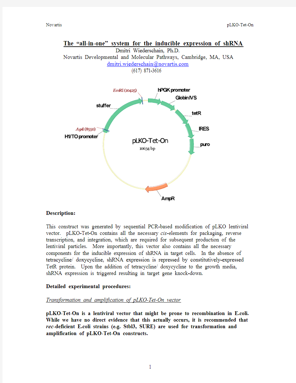
This construct was generated by sequential PCR-based modification of pLKO lentiviral vector. pLKO-Tet-On contains all the necessary cis-elements for packaging, reverse transcription, and integration, which are required for subsequent production of the lentiviral particles. More importantly, this vector also contains all the necessary components for the inducible expression of shRNA in target cells. In the absence of tetracycline/ doxycycline, shRNA expression is repressed by constitutively-expressed TetR protein. Upon the addition of tetracycline/ doxycycline to the growth media, shRNA expression is triggered resulting in target gene knock-down.
Detailed experimental procedures:
Transformation and amplification of pLKO-Tet-On vector
pLKO-Tet-On is a lentiviral vector that might be prone to recombination in E.coli. While we have no direct evidence that this actually occurs, it is recommended that rec-deficient E.coli strains (e.g. Stbl3, SURE) are used for transformation and amplification of pLKO-Tet-On constructs.
1. Transform pLKO-Tet-On vector into competent E.coli cells and streak out on
Amp plate.
2. Pick a single colony and expand into LB/Amp to obtain maxi DNA prep.
3. Confirm vector identity by restriction digest based on the sequence provided.
shRNA oligo design
Only 2 cloning sites are available in pLKO-Tet-On: AgeI and EcoRI. shRNA sequences from The RNAi Consortium collection (MISSION? shRNA, Sigma,
https://www.doczj.com/doc/3b1122225.html,), which utilizes constitutive pLKO vector, can be ordered as oligos and cloned into pLKO-Tet-On. In addition, a number of commercial software programs can be used to design and optimize shRNA sequences. The oligos are designed so that AgeI and EcoRI sites are mutated in the final construct to prevent oligo excision.
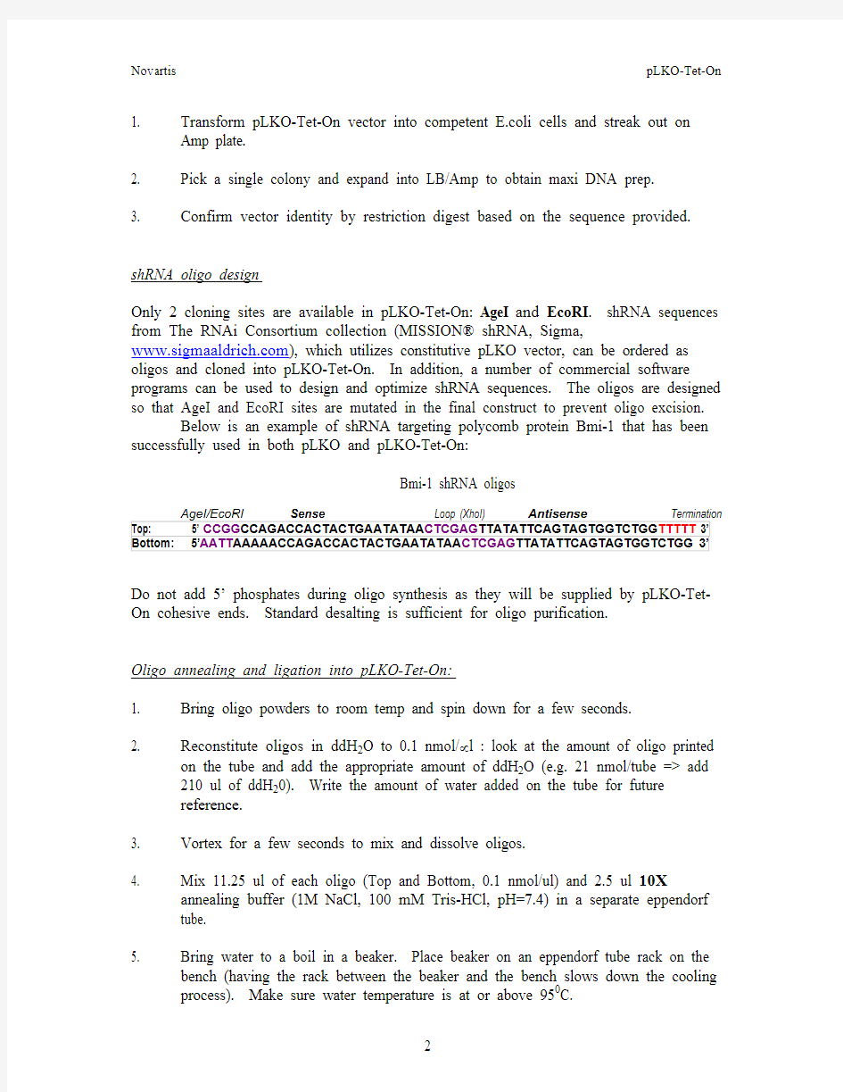
Below is an example of shRNA targeting polycomb protein Bmi-1 that has been successfully used in both pLKO and pLKO-Tet-On:
Bmi-1 shRNA oligos
AgeI/EcoRI Sense Loop (XhoI) Antisense Termination Top: 5’ CCGG CCAGACCACTACTGAATATAA CTCGAG TTATATTCAGTAGTGGTCTGG TTTTT 3’ Bottom: 5’AATT AAAAACCAGACCACTACTGAATATAA CTCGAG TTATATTCAGTAGTGGTCTGG 3’ Do not add 5’ phosphates during oligo synthesis as they will be supplied by pLKO-Tet-On cohesive ends. Standard desalting is sufficient for oligo purification.
Oligo annealing and ligation into pLKO-Tet-On:
1. Bring oligo powders to room temp and spin down for a few seconds.
2. Reconstitute oligos in ddH2O to 0.1 nmol/μl : look at the amount of oligo printed
on the tube and add the appropriate amount of ddH2O (e.g. 21 nmol/tube => add
210 ul of ddH20). Write the amount of water added on the tube for future
reference.
3. Vortex for a few seconds to mix and dissolve oligos.
4. Mix 11.25 ul of each oligo (Top and Bottom, 0.1 nmol/ul) and 2.5 ul 10X
annealing buffer (1M NaCl, 100 mM Tris-HCl, pH=7.4) in a separate eppendorf
tube.
5. Bring water to a boil in a beaker. Place beaker on an eppendorf tube rack on the
bench (having the rack between the beaker and the bench slows down the cooling process). Make sure water temperature is at or above 950C.
6. Place oligo mixtures in the water bath and allow to cool down naturally to app.
300C (2-3 hours).
7. Dilute 1 ul of the oligo mixture 1:400 in 0.5X annealing buffer (1 μl + 399 μl
0.5X buffer).
8. Annealed and diluted oligos can be stored at -200C.
9. Digest pLKO-Tet-On with AgeI and EcoRI and gel-purify the digested vector
(stuffer is released as a 1.8 kb fragment and should be discarded). Do not de-
phosphorylate pLKO-Tet-On after restriction digest with Age I/ EcoRI.
10. Set up ligation reaction as follows:
Oligo dilution (or 0.5X buffer as a negative control) 1 μl
Gel-purified digested pLKO-Tet-On (10-20 ng) 1 μl
10X ligase buffer 1 μl
T4 DNA ligase 1 μl
Water 6 μl
Total 10 μl
11. Always include “Vector only” negative control to monitor vector self-ligation.
12. Incubate at room temperature for 1-3 hours. Ligation reaction products can be
stored at -200C.
Transformation of ligation reaction product into E. coli:
1. Transform competent Stbl3 E. coli cells with 4 ul of the total ligation reaction
volume following established procedures (pre-chill ligation reaction on ice, add 4 ul of ligation reaction to E.coli, incubate for 20-30 min., heat shock for 30-45 sec.
at 420C, incubate on ice for 5 min, add 500 ul of SOC growth media, incubate at
370C while shaking for 30 min).
2. Plate 200 ul of E.coli cells onto pre-warmed LB/Carbenicillin-50μg/mL plates.
Screening for positive clones:
1. Pick individual colonies and inoculate miniprep cultures in 5-6 ml of LB/Amp
2. Isolate miniprep DNA using Qiagen kit. Elute DNA in 40-50 μl of pre-warmed
EB.
3. Set up XhoI restriction digest reaction using 4-5 μl of eluted DNA (30 μl total
reaction volume).
4. Resolve 20 μl of digestion reaction on 2% agarose gel.
5. Clones that contain shRNA oligos will yield 2 closely migrating bands at and
below 200 bp. In one example below, only clone #2 contains the shRNA insert.
Sequencing of pLKO-Tet-On shRNA constructs:
The following forward sequencing primer anneals upstream of the H1/TO promoter and has been successfully used to confirm the identity of shRNA constructs:
shRNA Seq primer: ggcagggatattcaccattatcgtttcaga
Example of Bmi-1 shRNA sequencing results:
…GTGAACGGATCTCGACGGTATCGATCACGAGACTAGCCTCGAGCGGCCGC
H1/TO promoter AATATTTGCATGTCGCTATGTGTTCTGGGAAATCACCATAAACGTGAAATCCC
Mutant AgeI TATCAGTGATAGAGACTTATAAGTTCCCTATCAGTGATAGAGACACC
shRNA
GG CCAGACCACTACTGAATATAACTCGAGTTATATTCAGTAGTGGTCTG Mutant EcoRI
GTTTTT AATTCTCGACCTCGAGACAAATGGCAGTATTCATCCA… Packaging of lentiviruses:
1. Day 1: Seed 4x10⁶ of 293T cells (passage 0-20) onto 10 cm collagen I- or poly-
D-lysine coated tissue culture plates in 10-15 ml of growth medium (DMEM/10% FBS).
2. Day 2: Change media to 6 ml of DMEM/10% FBS 1 hour before transfection.
Prepare transfection mixture of plasmid DNA and Fugene transfection
reagent (Roche) according to manufacturer’s instructions. A 1:3 ratio of
DNA : Fugene is used.
For 1 x 10 cm plate:
Add 36 ul of Fugene directly into 600 μl of OptiMEM. Incubate for 5
minutes at room temperature. If transfecting more than 1 plate, prepare
this mixture in bulk and then aliquot 636 μl/tube.
Mix the following plasmid DNA in 50 μl of OptiMEM:
pLKO-Tet-On shRNA 4 μg (must be high purity, ≥0.5μg/ul)
pLP1 3 μg
pLP2 3 μg
pLP/VSVG 2 μg
Add DNA to diluted Fugene reagent. Mix gently and incubate for 30
minutes. Add DNA/Fugene mixture (app. 700 μl) dropwise onto the cells.
3. Day 3: Change media to 6 ml DMEM/20% FBS per plate. Incubate for
additional 48 hours to generate lentivirus.
4. Day 5: Harvest viral supernatant (48 hours post-media change).
5. Spin down at 1500 rpm for 10 min.
6. Aliquot and freeze virus at -800C.
Titering of lentiviruses:
1. Plate 100,000 HCT116 cells (or other cell type)/well in a 6-well plate.
2. 24 hours later, change the media to growth media supplemented with 5-10 μg/ml
of polybrene (1 ml of media/well).
Note: Polybrene may be toxic to some cell lines. Determine proper
concentration of polybrene before transduction.
3. Add 50, 100, 200, 500 μl of viral supernatant to each well. Add 500 μl of media
to the “mock-transduced” and “no virus/ no puro” wells.
4. Incubate for 12-14 hours (overnight) in an incubator.
5. Change media to the relevant growth media supplemented with the desired
concentration of puromycin (1-3 μg/ml) in all viral infection wells and in “mock-transduced” well. Change media in “no virus/ no puro” well to fresh media with no puromycin.
6. Continually assess cell killing by puromycin in the “mock-transduced” well by
light microscopy. Change media as outlined in 5 every 48 hours.
7. Once all non-transduced cells have been eliminated by puro selection, stain the
dishes with crystal violet as follows:
- prepare crystal violet solution by dissolving 1 g of crystal violet powder
in 500 ml of buffered formalin (could be prepared as 4% formalin in PBS).
Shake well or stir until the powder has completely dissolved. Filter using
tissue culture filter unit.
- Aspirate media and rinse plates once with PBS.
- Apply 1 ml of stain to each well.
- Incubate for 20-30 min at room temperature; do not shake.
- Aspirate the stain and rinse several times with distilled water.
- Tap the plate upside down on a paper towel and air dry.
- Dishes can be scanned using regular document scanner.
8. To obtain a more quantitative assessment of viral titer, trypsinize and count the
cells in all wells (except “mock-transduced” where no cells should remain at this
point). The number of cells in the “no virus/ no puro” well is set to 100%.
Infectivity can be expressed as number of cells in each of virally-infected wells /
number of cells in “no virus/ no puro” well x 100%.
Example:
Number of cells in “no virus/ no puro” well – 10,000
Number of cells in “50 ul virus” well – 1,000
1,000/10,000 x 100% = 10% of cells were infected.
Perform the above calculation for each viral concentration used. Multiplicity of
infection (MOI) of 1 usually equals 50% infectivity.
Generation of stable inducible shRNA-expressing cell lines:
Use infection procedure above to infect target cell lines at optimum MOI. Generate stable pools of tranductants by selection with puromycin.
Important: Following transduction with lentiviral inducible shRNA, cells must be cultured in the growth media containing fetal bovine serum (FBS) approved for use with Tet-inducible systems (Clontech). Trace amounts of tetracycline in regular FBS might trigger shRNA expression.
Note: To avoid viral toxicity in your cell line, remove virus-containing media and replace with fresh media. Add puromycin-containing media 24 hours post media change to allow for sufficient expression of the puromycin resistance gene.
Induction of shRNA expression and gene knock-down:
Both tetracycline and doxycycline have been shown to induce shRNA expression using this system. Doxycycline is more stable in the culture medium (48 hours versus 24 hours for tetracycline) and appears to be more potent than tetracycline. 10-100 ng/ml of Dox has been shown to induce shRNA expression and target protein knock-down within 48 hours. In vivo, 1-2 mg/ml of doxycycline administered in 5% sucrose-containing drinking water can be used. Titration of the inducing agent (Dox or Tet), as well as determination of the optimum incubation period to achieve maximal target knock-down, is recommended for each individual target gene. It is also recommended to confirm TetR protein expression in the target cell line using anti-TetR antibody (TET02, MoBiTech/Boca Scientific).
PLEASE CITE THE FOLLOWING ARTICLES WHEN PUBLISHING DATA OBTAINED USING pLKO-Tet-On:
Wiederschain, D., Wee, S., Chen, L., Loo, A., Yang, G., Huang, A., Chen, Y., Caponigro, G., Yao, Y.-M., Lengauer, C., Sellers, W.R., and Benson, J.D. Single-vector inducible lentiviral RNAi system for oncology target validation, Cell Cycle. 8: 498-504, 2009.
Wee, S., Wiederschain, D., Maira, S.-M., Loo, A., Miller, C., deBeaumont, R., Stegmeier, F., Yao, Y.-M., and Lengauer, C. PTEN-deficient cancers depend on PIK3CB, Proc Natl Acad Sci U S A. 105: 13057-62, 2008.