237
Chapter 20
Cell Sensitivity Assays: The MTT Assay
Johan van Meerloo, Gertjan J.L. Kaspers, and Jacqueline Cloos
Abstract
The MTT (3-[4,5-dimethylthiazol-2-yl]-2,5 diphenyl tetrazolium bromide) assay is based on the conversion of MTT into formazan crystals by living cells, which determines mitochondrial activity. Since for most cell populations the total mitochondrial activity is related to the number of viable cells, this assay is broadly used to measure the in vitro cytotoxic effects of drugs on cell lines or primary patient cells. In this chapter the protocol of the assay is described including important considerations relevant for each step of the assay as well as its limitations and possible applications.
Key words: MTT, 3-[4,5-Dimethylthiazol-2-yl]-2,5 diphenyl tetrazolium bromide, Viability assay, IC 50, LC 50, Drug sensitivity assay, Cytotoxicity assay
The general purpose of the MTT (3-[4,5-dimethylthiazol-2-yl]-2,5
diphenyl tetrazolium bromide) assay is to measure viable cells in relatively high throughput (96-well plates) without the need for elaborate cell counting. Therefore the most common use is to determine cytotoxicity of several drugs at different concentra-tions. The principle of the MTT assay is that for most viable cells mitochondrial activity is constant and thereby an increase or decrease in the number of viable cells is linearly related to mito-chondrial activity. The mitochondrial activity of the cells is reflected by the conversion of the tetrazolium salt MTT into formazan crystals, which can be solubilised for homogenous measurement. Thus, any increase or decrease in viable cell number can be detected by measuring formazan concentration reflected in optical density (OD) using a plate reader at 540 and 720 nm. For drug sensitivity measurements the OD values of
1. I ntroduction
Ian A. Cree (ed.), Cancer Cell Culture: Methods and Protocols, Second Edition , Methods in Molecular Biology, vol. 731,DOI 10.1007/978-1-61779-080-5_20, ? Springer Science+Business Media, LLC 2011
238v an M eerloo, Kaspers, and Cloos
wells with cells incubated with drugs are compared to the OD of wells with cells not exposed to drugs.
The MTT assay is suitable for the measurement of drug sensitivity in established cell lines as well as primary cells. For dividing cells (usually cell lines) the decrease in cell number reflects cell growth inhibition and the drug sensitivity is then usually specified as the concentration of the drug that is required to achieve 50% growth inhibition as compared to the growth of the untreated control (50% inhibitory concentration, IC 50). For primary (nondividing) cells, drug sensitivity is measured as enhanced cell kill of treated cells as compared to the loss of cells already commonly seen in untreated cells (50% lethal con-centration, LC 50). For some cell types such as fresh acute myeloid leukemia (AML) cells the control median cell survival is 100%, which is no problem as long as results for treated cells are compared to the controls.
For primary pediatric acute lymphoblastic leukemia (ALL) cells, the in vitro MTT assay has been extensively applied to predict drug sensitivity in vivo. In these studies, a relatively good correlation was observed between in vitro sensitivity and clinical outcome (1–4). Despite these and other positive results, the MTT assay is not commonly used as predictive test for the clinic, but rather as an in vitro tool to determine potential antitumor activity of new drugs and/or combinations of drugs (5). In addition, the MTT assay is a suitable tool to study resistance to drugs (6, 7) (see Table 1 for applications).
In order to set up the MTT assay for cells and/or drugs that have not been tested before, several considerations are of crucial importance and will be discussed in this chapter.
Table 1
Possible applications for MTT assay
Application
References
239
Cell Sensitivity Assays: The MTT Assay Class 2B biocabinet suitable for drug experiments. Incubator
with 5% CO 2 at 37°C. Microplates – 96 well (Greiner Bio-one, Alphen a/d Rijn, The Netherlands). For suspension cells, we use round bottom wells or flat bottom wells, while for attached cells only flat bottoms can be used (see Subheading 3.1 for the different loading volume per plate) (see Note 1).
Dissolve 500 mg MTT powder (Sigma, St. Louis, USA) in 10 mL PBS. Stir with a magnetic stirrer for approximately 1 h in the dark. Filter sterilize the solution with a 0.22 m m filter (Millipore, Carrigtwohill, Ireland) and store in 10-mL aliquots at ?20°C.
Warning: MTT is toxic and harmful. MTT is light sensitive, hence protect from light.
To dissolve the formazan crystals, different solutions can be used such as methanol, ethanol, and DMSO. Our lab has the best experience with acidified isopropanol. To make this solution, add 50 mL 2 M HCl to 2.5 L isopropanol. Store the solution at least a month at room temperature before use. When the isopropanol is not acidified correctly the suspension will become cloudy. 1. Plateshaker (IKA schuttler MST4, Janke & Kunkerel, Staufen,
Germany). 2. Pipettes 0.001–1 mL, single channel and 0.01–0.3, multi-channel. 3. Class 2B hood. 4. Benchtop centrifuge.
5. Microplate reader (Anthos-Elisa-reader 2001, Labtec,
Heerhugowaard, The Netherlands). 6. O 2 incubator.
Generally cells are plated in triplicates to minimize the variability
of the results. The volume of cells depends on the type of plate used. For round bottom and flat bottom plates, 80 m L and 120 m L are used, respectively. Each plate should contain control wells (without drugs) and blank wells (without cells). For some drugs that also show absorbance at the given wavelenghts, an additional control is required of wells with medium (without cells) including
2. M aterials
2.1. C ells and Controls
2.2. Solutions and Solvents
2.2.1. M TT Solution
2.2.2. A cidified Isopropanol
2.3. E quipment
3. M ethods
3.1. P late Setup
240v an M eerloo, Kaspers, and Cloos
the range of drug used. The number of plates needed depends on the specific experiment. A common MTT assay experiment requires a testing plate for the OD of the drug, a testing plate to determine the growth curve for the starting amount of cells seeded per well, and a broad dilution range to determine the dilu-tion range for the experiments. However, for a standardized drug sensitivity assay of patient material, a single plate often suffices. For cell lines, it is recommended to include a day 0 plate in order to accurately determine the extent of growth (in the course of the experiment for the control cells). The outer wells are not used for the experiment due to evaporation and are filled with phos-phate buffered saline (PBS) to keep the evaporation of the plate to the minimum, some drugs can also have influence on neigh-bouring wells; this has to be investigated before starting the experiment (see Note 2).
There are two methods of drug exposure (1); if the drug is
unstable or cannot be stored in medium at ?20°C, the plates have to be prepared fresh by making the drug dilution stocks and adding the total amount of drugs in 20 m L for round bottom plates and 30 m L of drug solution to flat bottom wells to each well just before adding the cells. This renders the total volume of cell and drug suspension to 100 m L for round bottom and 150 m L for flat bottom 96-well plates. (2) For testing a stable drug, plates can be prepared with the 30 m L drug concentrations and can be stored in ?20°C for later use. When the cells are added to the wells containing the drug suspension, this will mix the drugs through the cells (see Note 2).
For primary leukemic cells, the plates are incubated for 4 days to determine the optimal effect for most standard drugs. For cell lines, the plates have to be incubated during log phase, which is usually 72–96 h in an incubator with 5% CO 2 at 37°C (see Note 1).After the appropriate incubation time, add 1:10 volume of MTT solution (5 mg/mL); e.g. 10 m L for round bottom and 15 m L for flat bottom 96-well plates. Unused MTT can be frozen and reused. Shake plates for 5 min on a plateshaker by slowly increas-ing the shaking speed to a maximum of 900 shakes/min. Then incubate the plate for another 4–6 h at 37°C in a CO 2 incubator, depending on the cell type.
150 m L of acidified isopropanol is added to each well and resuspended until all crystals have been dissolved. Mix each well thoroughly using a multichannel pipette. Between each row, rinse tips of multichannel pipette with isopropanol and discard the used isopropanol. Blow out tips thoroughly before mixing the next rows. Start with the control wells, before mixing the rows with drugs.
3.2. D rug Incubation
3.3. C ulture Period
3.4. M TT Incubation
3.5. Dissolution of Formazan Crystals
241
Cell Sensitivity Assays: The MTT Assay The OD is measured at 540 and 720 nm in order to get a more
exact measurement by correcting for background noise. The 720 nm OD background will be subtracted from the 540 nm OD total signal. The measured data are copied into an excel sheet and with the use of the following formula, the percentage of living cells can be determined: The average OD of the blank control wells (without cells and if the drug has no specific OD without drug as well) is subtracted from the average OD of the control wells (cells but no drugs) and the wells containing the drugs. Some drugs may interfere with the OD measurement and then the average OD of the wells containing drugs, but without cells is subtracted from the average OD of the control wells. The leukemic cell survival is calculated by: (OD treated well [?blank])/(mean OD control well [?blank]) × 100. The LC 50 (the drug concentration which results in 50% leukemic cell survival) can be calculated. For more reliable results the experiment should be done in triplicate and in duplicate in case of primary cells and when a range of drug concentrations is being tested.
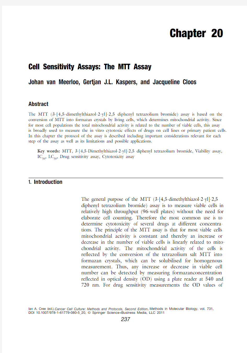
After adding acidified isopropanol and dissolving the formazan crystals thoroughly the plate has to rest for 10 min before measuring. In Fig. 1, a plate is shown after dissolving the formazan crystals with acidified isopropanol showing the blank culture medium wells, untreated cell control wells, and three drugs for which the cells are either sensitive (C), intermediate sensitive (E), and resistant (D) in these dose ranges. To be able
3.6. O D Measurement
3.7. E
xample Results
Fig. 1. A 96-well plate after formazan crystals are dissolved in acidified isopropanol.
(a ) Blanks control wells, (b ) untreated cell control wells, (c ) cell line with drug C with a dose–response curve from 100% cell death to no response on cell growth, (d ) cell line with drug D with a dose–response curve showing no growth inhibition, (e ) cell line with drug E with a dose–response curve with dose-dependent modest growth inhibition at high drug concentrations. Outer wells are not used because of possible evaporation.
242v an M eerloo, Kaspers, and Cloos
to determine IC 50 values of drugs D and E, different dose ranges are required.
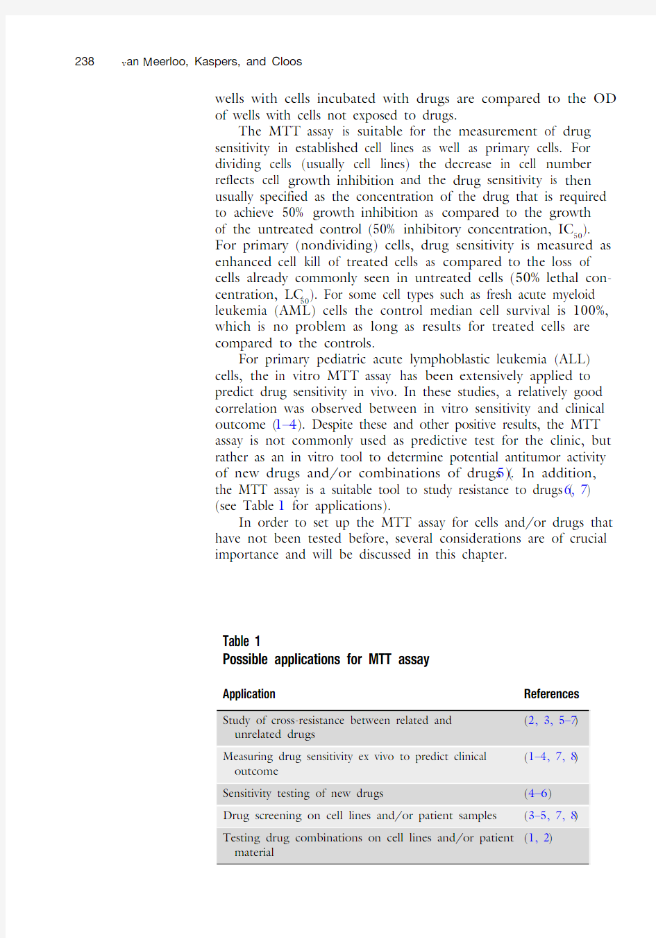
When measured in a plate reader, these measurements can then be used to determine the IC 50 for cell lines and LC 50 for patient material. The results can be plotted into a graph as shown in Fig. 2. The wells containing untreated cells are set as 100% and the dose–response curves of the three different drugs are depicted. It is clearly shown in the picture that for drugs D and E, the IC 50 is not reached.
1. The cell concentration that is plated may vary for different
cell lines and primary cells. For leukemic cell lines, we use concentrations in the range of 3.0–4.0 × 10³/well, while primary AML cells are plated in 0.08–0.12 × 10⁶ cells/well and primary ALL cells in 0.16 × 10⁶ cells/well. For cells that have not been used before in this assay, several cell concentration experiments have to be done before starting the drug experi-ments as recommended below. 2. Cells are plated in duplicate or triplicate, and in each plate,
4–6 control wells are included that contain the same amount of cells as the experimental wells and which will not be exposed to drugs. Additional 4–6 wells are needed for
4. N otes
% o f g r o w t h r e l a t i v e t o c o n t r o l
?10
0102030405060708090
100Fig. 2. Dose–response curve of cell lines C, D, and E. (C) cell line with drug C with a dose–response curve from control growth without drugs (set at 100%) to complete cell kill at the highest drug concentrations, (D) cell line with drug D with a dose–response curve showing no growth inhibition, (E) cell line with drug E with a dose–response curve showing dose-dependent modest growth inhibition (IC 50 is not reached).
243
Cell Sensitivity Assays: The MTT Assay
measuring the blanks, which contain the culture medium only. Some drugs can have an OD value of their own that can influence the measurement when diluted, and therefore a dilution of the drugs has to be measured without the adding cells. If there is a high difference in OD between the different drug dilutions, extra drug dilution control wells have to be added to the plate setup. The absorbance value for the blanks should be between 0.001 and 0.1 OD units as measured on a Microplate reader using filters for 540 and 720 nm. In addi-tion, the absorbance range for untreated cells should typically be between 0.75 and 1.25 OD units (see Notes 3 and 4). 3. To determine whether cells are suitable for use in the MTT assay, several parameters need to be verified:
(a) Linearity of viable cell number with OD level. To deter-
mine this, several cell concentrations are plated and
related to OD values. For cell types where the mitochon-
drial activity of cells is not constant, the MTT assay can-
not be used in to measure the influence of the drugs. (b) Cell concentrations. The optimal cell concentration to be
plated is dependent on the basic level of mitochondrial
activity and the rate of proliferation. In order to establish
this, several concentrations of cells should be plated in,
for instance, seven plates, and measured daily to deter-
mine the growth curve of the cell line to prevent over-
growth, which will influence the experiment. The starting
OD value of day 0 should not exceed 0.125. By con-
structing a growth curve, the logarithmic cell phase is
determined at which the cells duplicate. At a certain time
point the cell growth will plateau due to exhaustion of
the medium, contact inhibition, and exceeding of the
maximal OD value that can accurately be measured. The
most optimal concentration of plating is when cells have
almost no lag phase and the assay should not proceed
after the log phase.
(c) Cell culture. Cells should be cultured for a relevant time
period to be able to demonstrate the effect of the drug.
For cell lines this should only be during the log phase,
while for primary cells the MTT assay has to be completed
before all untreated cells are dead. Only for cell lines, a day
0 plate is used to precisely measure the activity of the
starting cell dilution at day 0 without the drugs’ effects.
Moreover, when using primary leukemic cells, in this
assay, the blast count at day 4 should be 370% leukemic
cells; this will be a blast count of 380–90% for ALL (7, 8)
and 370–75% for AML at day 0. Samples can be purified
with immunomagnetic beads directed against contaminat-
ing nonmalignant cells (8).
244v an M eerloo, Kaspers, and Cloos
4. Drug incubations.
(a) Control medium . In order to account for possible influences
of the dissolvent of the drugs on the background OD, the control medium should contain the concentrations of dis-solvent of drugs.(b) Stability of the drugs . To determine if cytostatic drugs
have to be added fresh or if the drugs can be stored in plates at ?20°C, drugs should be added into the 96-well plates and stored at ?20°C in advance of the experiments. Possible differences in the dose–response curves will imply whether the plates with drugs can be stored or that the drugs need to be added fresh at the day of plating.(c) Drug combinations of drug A + drug B can be measured
in several ways of which two are the most common (1) One fixed concentration of drug A can be used (usually in a concentration between the IC 20 and IC 50) in combi-nation with a dose range of drug B. (2) Mix both drugs at IC 50 as highest dose and then make a dose–response curve by diluting this mixture further to obtain a combined dose–response curve. Synergism or antago-nism can then be investigated, for instance, by the use of “calcusyn” software (Biosoft, Cambridge, UK) which can deal with both methods.(d) Reproducibility is important for an accurate measure-ment and so the use of at least a duplicate row setup of each drug, and if possible a triplicate row setup is pre-ferred within each plate. For cell lines we always perform the assay in triplicate while for primary material this is commonly not possible.(e) Plate set-up . It is our experience that some drugs can
affect surrounding cells possibly by aerosols; therefore, the wells with drugs have to be separated by PBS to make sure that the drugs will not affect the adjacent wells. The plate set-up also needs to include 4–6 control wells with the used culture medium, which are used to measure the background of culture medium.
Acknowledgments
This work was supported by KiKa “Stichting Kinderen Kankervrij” – Dutch Children Cancer-free foundation and VONK “VUmc Onderzoek Naar Kinderkanker” foundation.
245
Cell Sensitivity Assays: The MTT Assay References
1. Kaspers, G.J., Pieters, R., Van Zantwijk, CH.,
Van Wering, E.R., Van Der Does-Van Den Berg, A., Veerman, A.J. (1998) Prednisolone resistance in childhood acute lymphoblastic leukemia: vitro-vivo correlations and cross-resistance to other drugs. Blood.92, 259–266.
2. Salomons, G.S., Smets, L.A., Verwijs-Janssen,
M., Hart, A.A., Haarman, E.G., Kaspers, G.J., Wering, E.V., Der Does-Van Den Berg, A.V., Kamps, W.A. (1999) Bcl-2 family members in childhood acute lymphoblastic leukemia: rela-tionships with features at presentation, in vitro and in vivo drug response and long-term clini-cal outcome. Leukemia.13, 1574–1580.
3. Kaspers G.J., Veerman A.J., Pieters R, Van
Zantwijk CH., Smets L.A., Van Wering E.R., Van Der Does-Van Den Berg A. (1997) In vitro cellular drug resistance and prognosis in newly diagnosed childhood acute lymphoblas-tic leukemia. Blood. 90, 2723–2729.
4. Kaspers, G.J., Wijnands, J.J., Hartmann, R.,
Huismans, L., Loonen, A.H., Stackelberg, A., Henze, G., Pieters, R., H?hlen, K., Van Wering, E.R., Veerman, A.J. (2005) Immuno-phenotypic cell lineage and in vitro cellular drug resistance in childhood relapsed acute lymphoblastic leukaemia. Eur J Cancer.41, 1300–1303.
5. Hubeek, I., Peters, G.J., Broekhuizen, R.,
Zwaan, C.M., Kaaijk, P., van Wering, E.S.,
Gibson, B.E., Creutzig, U., Janka-Schaub,
G.E., den Boer, M.L., Pieters, R., Kaspers,
G.J. (2006) In vitro sensitivity and cross-resis-
tance to deoxynucleoside analogs in childhood acute leukemia. Haematologica.91, 17–23. 6. Oerlemans, R., Franke, N.E., Assaraf, Y.G.,
Cloos, J., van Zantwijk, I., Berkers, C.R., Scheffer, G.L., Debipersad, K., Vojtekova, K., Lemos, C., van der Heijden, J.W., Ylstra, B., Peters, G.J., Kaspers, G.J., Dijkmans, B.A., Scheper, R.J., Jansen, G. (2008) Molecular basis of bortezomib resistance: proteasome subunit beta5 (PSMB5) gene mutation and overexpression of PSMB5 protein. Blood.112, 2489–2499.
7. Klumper E., Pieters R., Veerman A.J.,
Huismans D.R., Loonen A.H., H?hlen K., Kaspers G.J., van Wering E.R., Hartmann R., Henze G. (1995) In vitro cellular drug resis-tance in children with relapsed/refractory acute lymphoblastic leukemia. Blood.86, 3861–3868.
8. Kaspers G.J., Veerman A.J., Pieters R,
Broekema G.J., Huismans D.R., Kazemier K.M., Loonen A.H., Rottier M.A., van Zantwijk CH., H?hlen K. (1994) Mononuclear cells contaminating acute lymphoblastic leu-kaemic samples tested for cellular drug resis-tance using the methyl-thiazol-tetrazolium assay. Br J Cancer. 70, 1047–1052.
细胞增殖及细胞活力检测方法 目前主要有两种用于检测细胞增殖能力的方法。一种是直接的方法,通过直接测定进行分裂的细胞数来评价细胞的增殖能力。另一种是间接的方法,即细胞活力(cell viability)检测方法,通过检测样品中健康细胞的数目来评价细胞的增殖能力。显然,细胞活力检测法并不能最终证明检测样品中的细胞是否在增殖。如细胞在某一培养条件下会自发启动凋亡程序,但药物的干扰可抑制凋亡的发生;这时若采用细胞活力检测法,显然可以区分两种条件下的细胞数量,但我们并不能从药物干扰组细胞数大于对照组的事实说明药物可促进细胞增殖的结论。所以最直接的证据应该采用方法一。 用于检测细胞增殖能力最经典的方法是用氚标记的胸腺嘧啶核苷处理细胞,再检测DNA链中氚含量。若细胞具有增殖能力,DNA合成过程中将会采用氚标记的胸腺嘧啶核苷作为合成原料,因此检测细胞DNA链内标记核苷酸的量可判断细胞是否进行DNA 的合成。 但更为常用的方法是BrdU检测法。用BrdU预处理的细胞中,BrdU可代替胸腺嘧啶核苷插入复制的DNA双链中,而且这种置换可以稳定存在,并带到子代细胞中。细胞经过固定和变性处理后,可用免疫学方法检测DNA中BrdU的含量(如采用鼠抗BrdU单克隆抗体特异识别BrdU,再采用辣根过氧化酶标记的山羊抗鼠IgG二抗标记,最后用比色法或荧光的方法进行定量测定),从而判断细胞的增殖能力。Calbiochem/EMD公司提供一种BrdU检测试剂盒,以微孔板的形式,合并所有清洗、固定、变性的步骤以单一试剂当中。比色检测在一抗二抗标记后在450nm下读数,
所有操作在3小时内结束。而且该试剂盒的灵敏度与市场上其他同类产品相比是最强的。1000个细胞以上水平的检测只需用BrdU预孵育2小时,100个细胞则采用过夜预孵育,即可检测细胞的增殖能力。 BrdU法的一个缺点是需要固定和变性等破坏DNA的处理。有些情况下,研究者可能希望在测定细胞增殖能力的同时检测细胞的总DNA含量,然而,在变性条件下,DNA 的双链结构将被破坏,DAPI和Hoechest 33342等核酸标记探针就不再能识别DNA,因而也无法估计DNA总量。Molecular Probes公司的Click-iT EdU检测试剂盒可以解决这个问题。这种方法不需要变性步骤,因为荧光探针标记的叠氮化物小分子,而不是庞大的抗体分子,可以很轻易的识别并结合未变性DNA双链中的EdU分子。采用BrdU方法时,你必须非常小心的去对DNA进行变性,才能一方面使BrdU抗体进入细胞,另一方面又保留足够的双链DNA分子来进行细胞周期的分析。有了EdU 后则不同,由于你不再需要变性,这一切都很简单了;另外,常常用于DNA变性的HCl,可能破细胞内坏蛋白的抗原识别位点,因而限制了BrdU检测法中同时检测其他蛋白的应用,但这种情况在EdU法中不存在。 图1:EdU及BrdU原理示意图(摘至invitrogen说明书) 在一些情况下,细胞活力的检测相当于细胞增殖能力的测定。用于细胞活力检测的方法又很多,这些方法主要采用特殊的试剂来测定细胞的代谢活力,Alamar Blue,MTT及其他四唑盐。它们通过检测细胞的氧化还原活性来检测细胞增殖能力,所以这是一种间接的方法。 Calbiochem的快速细胞增殖试剂盒,或者,严格来说,叫细胞活力试剂盒,采用一
细胞增殖及细胞活力检 测方法 Document number:NOCG-YUNOO-BUYTT-UU986-1986UT
细胞增殖及细胞活力检测方法 目前主要有两种用于检测细胞增殖能力的方法。一种是直接的方法,通过直接测定进行分裂的细胞数来评价细胞的增殖能力。另一种是间接的方法,即细胞活力(cell viability)检测方法,通过检测样品中健康细胞的数目来评价细胞的增殖能力。显然,细胞活力检测法并不能最终证明检测样品中的细胞是否在增殖。如细胞在某一培养条件下会自发启动凋亡程序,但药物的干扰可抑制凋亡的发生;这时若采用细胞活力检测法,显然可以区分两种条件下的细胞数量,但我们并不能从药物干扰组细胞数大于对照组的事实说明药物可促进细胞增殖的结论。所以最直接的证据应该采用方法一。 用于检测细胞增殖能力最经典的方法是用氚标记的胸腺嘧啶核苷处理细胞,再检测DNA 链中氚含量。若细胞具有增殖能力,DNA合成过程中将会采用氚标记的胸腺嘧啶核苷作为合成原料,因此检测细胞DNA链内标记核苷酸的量可判断细胞是否进行DNA的合成。 但更为常用的方法是BrdU检测法。用BrdU预处理的细胞中,BrdU可代替胸腺嘧啶核苷插入复制的DNA双链中,而且这种置换可以稳定存在,并带到子代细胞中。细胞经过固定和变性处理后,可用免疫学方法检测DNA中BrdU的含量(如采用鼠抗BrdU单克隆抗体特异识别BrdU,再采用辣根过氧化酶标记的山羊抗鼠IgG二抗标记,最后用比色法或荧光的方法进行定量测定),从而判断细胞的增殖能力。 Calbiochem/EMD公司提供一种BrdU检测试剂盒,以微孔板的形式,合并所有清洗、固定、变性的步骤以单一试剂当中。比色检测在一抗二抗标记后在450nm下读数,所有操作在3小时内结束。而且该试剂盒的灵敏度与市场上其他同类产品相比是最强的。1000个细胞以上水平的检测只需用BrdU预孵育2小时,100个细胞则采用过夜预孵育,即可检测细胞的增殖能力。 BrdU法的一个缺点是需要固定和变性等破坏DNA的处理。有些情况下,研究者可能希望在测定细胞增殖能力的同时检测细胞的总DNA含量,然而,在变性条件下,DNA的双链结构将被破坏,DAPI和Hoechest 33342等核酸标记探针就不再能识别DNA,因而也无法估计DNA总量。Molecular Probes公司的Click-iT EdU检测试剂盒可以解决这个问题。这种方法不需要变性步骤,因为荧光探针标记的叠氮化物小分子,而不是庞大的抗体分子,可以很轻易的识别并结合未变性DNA双链中的EdU分子。 采用BrdU方法时,你必须非常小心的去对DNA进行变性,才能一方面使BrdU抗体进入细胞,另一方面又保留足够的双链DNA分子来进行细胞周期的分析。有了EdU后则不同,由于你不再需要变性,这一切都很简单了;另外,常常用于DNA变性的HCl,可能破细胞内坏蛋白的抗原识别位点,因而限制了BrdU检测法中同时检测其他蛋白的应用,但这种情况在EdU法中不存在。
细胞活性测定方法 细胞活性指标通常包括细胞膜对核酸染料的通透性,代谢活性,膜电位等。核酸染料有多种,如EB带有单个自由正电荷,能通过完整细胞膜。而PI,TO—PRO—1,TO-PRO-3等等带一个有四铵基团和两个或两个以上正电荷的染料是不能通过完整细胞膜进入细胞内的。因而吸收了这些多电荷染料的细胞被认为是非活性的。另外,一些酸性染料,如上面提到的台盼兰,曙红等都是膜非通透性。 代谢活性是另一重要指标,它通过细胞内的酶的活性来判定。使用细胞某种酶的底物,它能通过(或是不能通过)完整细胞膜,在细胞内被酶切而产生荧光性,膜不通透性产物,能在细胞膜完好的细胞内存留,在细胞膜不完整的细胞内散失很快。通过检测荧光强度就可知细胞代谢活性。FDA(fluorescein diacetate)和CTC(5-cyano-2,3-ditolyl tetrazolium chloride)是常用的两种底物。前者虽然透过细胞膜的速度较慢,但它的产物基本不往外通透。后者经细胞内脱氢酶催化而具有荧光性,能提供细胞呼吸代谢系统活性和细胞膜完整性信息。 正常细胞的细胞膜两侧维持着一个胞内为负的膜电位为梯度,带正电的亲脂性染料,如Cyanines类能因电梯度而通过细胞的脂双层膜聚积在活细胞内,带负电的亲脂性染料如oxonols会被排除在外。不再维持着膜电位梯度的细胞里,则会吸收更多Oxonols类染料。 用流式细胞仪测量的方法优点是,灵敏,荧光强度精确定量,快速高通量的检测逐个细胞,可同时检测细胞的多个活性指标,提高可信度,结果具有统计意义。缺点是,实验较MTT等复杂,费用较高。 常用的细胞活性测定方法有台盼蓝染色法、克隆(集落)形成法、3H放射性同位素掺入法、MTT法等。其中MTT法以其快速简便,不需要特殊检测仪器、无放射性同位素、适合大批量检测的特点而得到广泛的应用。但MTT法形成的Formazan为水不溶性的,需要加有机溶剂溶解,由于在去上清操作时会有可能带走小部分的Formazan,故有时重复性略差。为了解决这个问题,研究人员又开发了很多种水溶性的四氮唑盐类:如XTT、CCK-8(WST-8)等。 1.MTT法 MTT:化学名:3-(4,5-二甲基噻唑-2)-2,5-二苯基四氮唑溴盐,商品名:噻唑蓝。检测原理为活细胞线粒体中的琥珀酸脱氢酶能使外源性MTT还原为水不溶性的蓝紫色结晶甲臜(Formazan)并沉积在细胞中,而死细胞无此功能。二甲基亚砜(DMSO)能溶解细胞中的甲臜,用酶联免疫检测仪在490nm波长处测定其光吸收值,可间接反映活细胞数量。在一定细胞数范围内,MTT结晶形成的量与细胞数成正比。该方法已广泛用于一些生物活性因子的活性检测、大规模的抗肿瘤药物筛选、细胞毒性试验以及肿瘤放射敏感性测定等。它的特点是灵敏度高、经济。 缺点:由于MTT经还原所产生的甲臜产物不溶于水,需被溶解后才能检测。这不仅使工作量增加,也会对实验结果的准确性产生影响,而且溶解甲臜的有机溶剂对实验者也有损害。
细胞活性测定方法介绍和使用探讨 细胞活性测定方法有台盼蓝染色法、克隆(集落)形成法、3H放射性同位素掺入法、MTT法等。其中MTT法以其快速简便,不需要特殊检测仪器、无放射性同位素、适合大批量检测的特点而得到广泛的应用。但MTT法形成的Formazan为水不溶性的,需要加有机溶剂溶解,由于在去上清操作时会有可能带走小部分的Formazan,故有时重复性略差。为了解决这个问题,研究人员又开发了很多种水溶性的四氮唑盐类:如XTT、CCK-8(WST-8)等。 现就这三种四氮唑盐类方法作一个简单介绍: 1、 MTT 法 MTT:化学名:3-(4,5-二甲基噻唑-2)-2,5-二苯基四氮唑溴盐,商品名:噻唑蓝。检测原理为活细胞线粒体中的琥珀酸脱氢酶能使外源性MTT还原为水不溶性的蓝紫色结晶甲瓒(Formazan)并沉积在细胞中,而死细胞无此功能。二甲基亚砜(DMSO)能溶解细胞中的甲瓒,用酶联免疫检测仪在490nm波长处测定其光吸收值,可间接反映活细胞数量。在一定细胞数范围内,MTT结晶形成的量与细胞数成正比。该方法已广泛用于一些生物活性因子的活性检测、大规模的抗肿瘤药物筛选、细胞毒性试验以及肿瘤放射敏感性测定等。它的特点是灵敏度高、经济。 缺点:由于MTT经还原所产生的甲瓒产物不溶于水,需被溶解后才能检测。这不仅使工作量增加,也会对实验结果的准确性产生影响,而且溶解甲瓒的有机溶剂对实验者也有损害。 2、 XTT 法 XTT,化学名: 2,3-bis(2-methoxy-4-nitro-5-sulfophenyl)-5-[(phenylamino)carbonyl]-2H-tetrazolium hydroxide,作为线粒体脱氢酶的作用底物,被活细胞还原成水溶性的橙黄色甲瓒产物。当XTT与电子偶合剂(例如PMS)联合应用时,其所产生的水溶性的甲瓒产物的吸光度与活细胞的数量成正比。 优点:1、使用方便,省去了洗涤细胞;2、检测快速;3、灵敏度高,甚至可以测定较低细胞密度;4、重复性优于MTT。 缺点:XTT水溶液不稳定,需要低温保存或现配现用。
1、细胞活性的鉴定:死细胞对许多抗体均有很强的非特异性染色,这就使样本细胞活性检测变得非常重要,尤其是经过长时间运输和储存的样本。检测的方法通常有两种:(1)实时的流式检测:利用荧光染料PI、7-AAD或EMA 进行死细胞染色,而活细胞拒染这些染料。此方法的优势是细胞表面标志和活性分析可同时进行。尤其适用于高度坏死的样本。7-AAD最常用,因为在488nm激发下,其最大发射光在670nm左右,适合与FITC或PE进行多色标记。但随着时间延长,7-AAD会在固定的细胞群体重新分配,死活细胞的区分变得困难。因此,对于染色并在固定后12小时以上分析的标本,最好用EMA。EMA与死细胞DNA稳定的共价结合保证了长时间固定后仍能很好的区分固定前的死活状态。(2)手工检测:使用Trypanblue或其他细胞活性染料。(3)使用专门的仪器进行检测。如Vi-cell。 2、7-AAD (7-amino-actinomycin D)是一种核酸染料,在流式细胞术中用于鉴定死细胞,这种作用与PI相似。它不能通过正常质膜,随着细胞凋亡、细胞死亡过程,质膜对7-AAD的通透性逐渐增加,结合细胞凋亡中DNA的有控降解,在合适波长激发光的激发下可发出明亮的红色荧光,通过7-AAD标记DNA的强弱,将细胞分为三群:7-AAD 强为死亡细胞,7-AAD弱是凋亡细胞,7-AAD-为正常活力细胞。7-AAD同PI 有着相似的荧光特性,但其发射波谱较PI窄,PI检测时同时占据FL-2和FL-3通道,而7-AAD只占FL-3通道,对其他检测通道的干扰更小,在多色荧光分析中是PI 的最佳替代品,可与Annexin V-EGFP/FITC/PE联合使用。 3、Annexin V-PE/7-AAD细胞凋亡检测试剂盒是用红色荧光染料PE(Phycoerythrin)标记的AnnexinV作为探针,来检测细胞早期凋亡的发生,可用荧光显微镜、流式细胞仪或其他荧光检测设备进行检测。 其检测原理为:在正常的活细胞中,磷脂酰丝氨酸(phosphotidylserine,PS)位于细胞膜的内侧,但在早期凋亡的细胞中,PS 从细胞膜的内侧翻转到细胞膜的表面,暴露在细胞外环境中。Annexin-Ⅴ(膜联蛋白-V)是一种分子量为35-36KD的Ca2+依赖性磷脂结合蛋白,能与PS高亲和力结合。可通过细胞外侧暴露的磷脂酰丝氨酸与凋亡早期细胞的胞膜结合。因此Annexin V是检测细胞早期凋亡的灵敏指标。 另外,本试剂盒中提供的7-AAD可用来区分存活的早期细胞和坏死或晚期凋亡细胞。7-AAD是一种核酸染料,同PI 有着相似的荧光特性,但其发射光谱较PI窄,对其他检测通道的干扰更小,在多色荧光分析中是PI的最佳替代品,可与Annexin V联合使用。该染料不能透过正常细胞或早期凋亡细胞的完整的细胞膜,但可穿透晚期凋亡细胞或者坏死细胞并与其内的DNA结合。因此将AnnexinV-PE与7-AAD联合使用时,7-AAD则被排除在活细胞(AnnexinV-/7-AAD-)和早期凋亡细胞(AnnexinV+/7-AAD-)之外,而晚期凋亡细胞和坏死细胞同时被AnnexinV-PE 和7-AAD结合染色呈现双阳性(AnnexinV+/7-AAD+),可以将凋亡早期的细胞和晚期的细胞以及死细胞区分开来。。 操作方法 样品染色 1)悬浮细胞:300g,4℃离心5 min收集细胞。 贴壁细胞:用不含EDTA的胰酶消化后,300g,4℃离心5 min收集细胞。胰酶消化时间不宜过长,以防引起假阳性。2)用4℃预冷的PBS洗涤细胞2次,每次均需300g,4℃离心5 min。 3)加入250μl 1×Binding Buffer重悬细胞,调节其浓度为1×10⁶细胞/ml。 4)取100 μl细胞悬液于5ml流式管中,加入5μl Annexin V/PE和10 μl7-AAD,轻轻混匀。 5)避光、室温反应15 min。 6)加入400μl1×Binding Buffer,混匀,样品在1小时内检测。 4、CFSE (carboxyfluoresceindiacetate, succinimidyl ester 羧基荧光素双乙酸盐,琥珀酰亚胺酯) 被动扩散进入细胞,其本身是无色无荧光的,被细胞内的酯酶分解后产生高强度的绿色荧光,定位于细胞膜、细胞质和细胞核,在细胞核的荧光染色最强,该荧光产物与胞内的氨基结合长时间留存于细胞内并可被乙醛固定。未结合的试剂与副产物扩散至胞外基质中,被洗去。 传代或胚胎分裂分析,CFSE与赖氨酸侧链或其他氨基基团反应,共价偶联至细胞内和细胞表面的蛋白,细胞分裂时被平均分配至子代细胞,荧光强度降到母代细胞的一半。适合用于胞内示踪,在细胞分裂或融合后分配至子代细胞中,并不会被转移至邻近的细胞。在小鼠淋巴细胞迁移实验中,CFSE注射进入小鼠体内后,标记的淋巴细胞的检测可长达8周。肝内注射荧光显微镜定位检测可长达20天。
MTT检测细胞活性的操作方法 一、MTT是什么 MTT是一种粉末状化学试剂,全称为3-(4,5)-dimethylthiahiazo (-z-y1)-3,5-di-phenytetrazoliumromide,汉语化学名为3-(4,5-二甲基噻唑-2)-2,5-二苯基四氮唑溴盐,商品名:噻唑蓝。是一种黄颜色的染料。 二、MTT法用来做什么 简单地说:是一种检测细胞存活和生长的方法。 MTT主要有两个用途 1.药物(也包括其他处理方式如放射线照射)对体外培养的细胞毒性的测定; 2.细胞增殖及细胞活性测定。 三、为何MTT可以用来做上述工作 检测原理为活细胞线粒体中的琥珀酸脱氢酶能使外源性MTT还原为水不溶性的蓝紫色结晶甲瓒(Formazan)并沉积在细胞中,而死细胞无此功能。二甲基亚砜(DMSO)能溶解细胞中的甲瓒,用酶标仪在490nm波长处测定其光吸收值,在一定细胞数范围内,MTT结晶形成的量与细胞数成正比。根据测得的吸光度值(OD值),来判断活细胞数量,OD值越大,细胞活性越强(如果是测药物毒性,则表示药物毒性越小)。 四、实验所需材料 1.MTT 溶液的配制通常MTT 配成的终浓度为5mg/ml,须用PBS或生理盐水做溶剂。 市面上一般MTT的包装为100mg,250mg或1g 1.1对于100mg这样的小包装,厂家都是将MTT放入小管中的,个人建议不要再用天平称量分装,而应该一次性将其全配制成溶液,如100mg用20mlPBS来溶解。具体做法:预先在50ml离心管(没有的话,可用培养瓶替代)加入20ml PBS,从中先吸取500-1000ul PBS装入含MTT的小管中,吹打若干次后将其移入50ml离心管,然后再混匀。可以重复几次,以使小管中的MTT不残留于管内。将MTT完全混匀后,用0.22μm滤膜过滤以除去溶液里的细菌,分装避光(避光袋或是黑纸、锡箔纸包住)可长期保存于-20度。按细胞培养板每孔需加10ul计算,一般每96孔板约需1ml,所以分装时可考虑每管分装1ml。 1.2对于大包装,可按上述方法称取一部分溶解,也有人将粉剂分装在EP管里,用的时候现配,直接往培养板中加。 注意事项: 1. 在配制和保存的过程中,容器最好用铝箔纸包住。 配成的MTT需要无菌,MTT对菌很敏感 MTT一般最好现用现配,过滤后4度避光保存两周内(个人曾做过4度避光保存4周的MTT溶液,效果仍然不错)有效,或配制成5mg/ml保存在-20度长期保存,避免反复冻融,最好小剂量分装,用避光袋或是黑纸、锡箔纸包住避光以免分解。当MTT变为灰绿色时就绝对不能再用。 MTT有致癌性,用的时候小心,有条件最好带那种透明的簿膜手套. 2.MTT甲瓒溶解液 2.1二甲基亚砜DMSO,可以直接溶解,无需配制,使用方便,溶解速度快,但对人体毒性较大,且需去除原培养液,在去除培养液的过程中,可能会把结晶去掉,导致结果不稳定。 2.2三联溶解液:SDS10g,异丁醇5ml,10M HCl 0.1ml 用双蒸水溶解配成100ml溶液,该溶解液不需去除原培养基,但溶解较慢。 该溶解液因含有SDS,在低温保存的时候易产生结晶,因此在用之前必须提前几小时
ROS活性氧检测试剂盒-DCFHDA法 产品组成: 产品编号BB-47053 规格200-1000T DCFHDA100ul 活性氧阳性对照诱导剂1000ul 储存条件: -20℃保存。 有效期: 一年。 注意事项: ● 本试剂盒仅供科学研究使用,不可用于诊断或治疗。 ● 螺旋盖微量试剂管装的试剂在开盖前请短暂离心,将盖和管内壁上的液体离心至管 底,避免开盖时试剂损失。 ● 禁止与其他品牌的试剂混用,否则会影响使用效果。 ● 样品或试剂被细菌或真菌污染或试剂交叉污染可能会导致错误的结果。 ● 最好使用一次性吸头、管、瓶或玻璃器皿,可重复使用的玻璃器皿必须在使用前清 洗并彻底清除残留清洁剂。 ● 避免皮肤或粘膜与试剂接触。 产品简介: 活性氧(Reactive oxygen species, ROS) 包括超氧自由基、过氧化氢、及其下游产物过氧化物和羟化物等,参与细胞生长增殖、发育分化、衰老和凋亡以及许多生理和病理过程。 贝博活性氧检测试剂盒是一种利用荧光探针DCFH-DA进行活性氧检测的试剂盒。DCFH-DA本身没有荧光,可以自由穿过细胞膜,进入细胞内后,可以被细胞内的酯酶水解生成DCFH。而DCFH不能通透细胞膜,从而使探针很容易被标记到细胞内。在活性氧存在的条件下,DCFH被氧化生成荧光物质DCF,绿色荧光强度与细胞内活性氧水平成正比,检测DCF的荧光就可以知道细胞内活性氧的水平。 在激发波长502 nm,发射波长530 nm附近,使用荧光显微镜、激光共聚焦显微镜、荧光分光光度计、荧光酶标仪、流式细胞仪等检测DCF 荧光,从而测定细胞内活性氧水平。 本试剂盒可以用于各种真核培养细胞的检测。本试剂盒提供了活性氧阳性对照试剂,以便于活性氧的检测。 产品特点: ● 使用方便:可用激光共聚焦显微镜直接观察、荧光分光光度计、荧光酶标仪或流式细胞仪检测; ● 背景低,灵敏度高; ● 线性范围宽,使用方便。
检测细胞活性的操作方法 一、是什么 是一种粉末状化学试剂,全称为() (),汉语化学名为(,二甲基噻唑),二苯基四氮唑溴盐,商品名:噻唑蓝。是一种黄颜色的染料。 二、法用来做什么 简单地说:是一种检测细胞存活和生长的方法。 主要有两个用途 .药物(也包括其他处理方式如放射线照射)对体外培养的细胞毒性的测定; .细胞增殖及细胞活性测定。 三、为何可以用来做上述工作 检测原理为活细胞线粒体中的琥珀酸脱氢酶能使外源性还原为水不溶性的蓝紫色结晶甲瓒()并沉积在细胞中,而死细胞无此功能。二甲基亚砜()能溶解细胞中的甲瓒,用酶标仪在波长处测定其光吸收值,在一定细胞数范围内,结晶形成的量与细胞数成正比。根据测得的吸光度值(值),来判断活细胞数量,值越大,细胞活性越强(如果是测药物毒性,则表示药物毒性越小)。 四、实验所需材料 .溶液的配制通常配成的终浓度为,须用或生理盐水做溶剂。 市面上一般的包装为或 对于这样的小包装,厂家都是将放入小管中的,个人建议不要再用天平称量分装,而应该一次性将其全配制成溶液,如用来溶解。具体做法:预先在离心管(没有的话,可用培养瓶替代)加入,从中先吸取装入含的小管中,吹打若干次后将其移入离心管,然后再混匀。可以重复几次,以使小管中的不残留于管内。将完全混匀后,用μ滤膜过滤以除去溶液里的细菌,分装避光(避光袋或是黑纸、锡箔纸包住)可长期保存于度。按细胞培养板每孔需加计算,一般每孔板约需,所以分装时可考虑每管分装。 对于大包装,可按上述方法称取一部分溶解,也有人将粉剂分装在管里,用的时候现配,直接往培养板中加。 注意事项: . 在配制和保存的过程中,容器最好用铝箔纸包住。 配成的需要无菌,对菌很敏感 一般最好现用现配,过滤后度避光保存两周内(个人曾做过度避光保存周的溶液,效果仍然不错)有效,或配制成保存在度长期保存,避免反复冻融,最好小剂量分装,用避光袋或是黑纸、锡箔纸包住避光以免分解。当变为灰绿色时就绝对不能再用。 有致癌性,用的时候小心,有条件最好带那种透明的簿膜手套. .甲瓒溶解液 二甲基亚砜,可以直接溶解,无需配制,使用方便,溶解速度快,但对人体毒性较大,且需去除原培养液,在去除培养液的过程中,可能会把结晶去掉,导致结果不稳定。 三联溶解液:,异丁醇,用双蒸水溶解配成溶液,该溶解液不需去除原培养基,但溶解较慢。 该溶解液因含有,在低温保存的时候易产生结晶,因此在用之前必须提前几小时拿至室温,将结晶全部溶解后再使用。
MTT法测定细胞相对数和相对活力 一、原理 噻唑兰,简称MTT,可透过细胞膜进入细胞内,活细胞线粒体中的琥珀脱氢酶能使外源性MTT还原为难溶于水的蓝紫色的Formazan结晶并沉积在细胞中,结晶物能被二甲基亚砜(DMSO)溶解,用酶联免疫检测仪在490nm波长处测定其光吸收值,可间接反映细胞数量。 二、试剂材料准备与实验仪器 1)对数生长期细胞 2)受试因素(药物) 3)MTT:以PBS配制成5mg /ml,抽滤除菌,保存在4?C 4)DMSO(二甲基亚砜) 5)96孔板 6)酶联免疫检测仪 7)细胞培养箱 三、实验步骤(适用于贴壁细胞) 1)收集对数期细胞,调整细胞悬液浓度,分于96孔板,1×104/孔,细胞浓度可以调整。2)置37℃、5%CO2温箱培养使细胞贴壁。 3)加入不同浓度的药物,如1、5、10、40、50、80、160、320mg/ml的药物,时间依据实验需要,一般3天。 4)小心吸去上清,PBS轻轻洗涤,再次离心,弃上清。 5)每孔加入180 ul新鲜RPMI 1640培养液,再加入20 ul MTT溶液(5 mg/ml,即0.5%MTT),继续培养4 h。
6)终止培养(可离心,1000 rpm,10 min),小心吸去孔内培养液。 7)每孔加入150 ul二甲基亚砜(也可以用酸化异丙醇,10%SDS代替),置摇床上低速振荡10 min,使结晶物充分溶解。在酶联免疫检测仪490 nm处测量各孔的吸光值。8)同时设置调零孔(培养基、MTT、二甲基亚砜),对照孔(细胞、相同浓度的药物溶解介质、培养液、MTT、二甲基亚砜),每组设定3复孔。 三、结果统计学处理 所有数值以x±s表示,应用SPSS软件进行方差分析,p<0.05时为相差显著,p<0.01时为相差非常显著。可以以时间为横轴,光吸收值为纵轴绘制细胞生线,专门公式求IC50。或计算抑制率。 四、注意事项与常见问题 1)实验时应设置调零孔,对照孔,加药孔。调零孔加培养基、MTT、二甲基亚砜。对照孔和加药孔都要加细胞、培养液、MTT、二甲基亚砜,不同的是对照加溶解药物的介质,而加药组加入不同浓度的药物。 2)每孔中的细胞数可以根据细胞生长的速度调整,并进行预实验调整浓度,太多敏感性降低,太少观察不到差异。 3)贴壁细胞加MTT前吸掉培养液,悬浮细胞可以不吸除培养液,再加入0.5%MTT,量为20ul/孔。 4)如果不使用96孔板,培养基超过100 ml,MTT按照10%的比例加入。 5)MTT一般4度保存两周,注意避光保存,或配制成5mg/ml保存在-20度长期保存,避免反复冻融,最好小剂量分装,用避光袋或是黑纸、锡箔纸包住避光以免分解,配制完后可过滤一下。
CCK-8细菌活性检测试剂盒 产品组成: 产品编号BB-4221-1 BB-4221-2 BB-4221-3 规格250T 500T 1000T CCK-8细菌检测试剂 2.5ml 5ml 10ml 产品简介: 贝博细菌活性检测试剂盒(CCK-8法) 是应用新型的水溶性四唑盐2-(2-甲氧基-4-硝苯基)-3-(4-硝苯基)-5-(2,4-二磺基苯)-2H-四唑单钠盐快速高灵敏度检测细菌活性的比色检测产品。 本四唑盐在电子载体存在的情况下能够被细菌的脱氢酶还原成橙黄色的水溶性的Formazan,生成的Formazan量与细菌的活性成正比。细菌活性越强,则颜色越深;细菌活性越弱,则颜色越浅。对于同样的细菌,颜色的深浅和细菌活性呈线性关系。 CCK-8溶液可以直接加入到细菌样品中,不需要预配各种成分,CCK-8溶液很稳定,对细菌没有毒性,可以长时间培养细菌。 图一:CCK-8细菌活性检测原理 使用方法: 1. 制备适当浓度微生物细胞的悬液,在96孔板内每孔接种190μl悬液。 2. 每孔加入10μl的染色溶液。 3. 将培养板在培养箱中培养(37℃或合适的温度)。 4. 用酶标仪测定在450nm处的吸光度。 细胞活力计算: 各重复孔的OD值取平均数。 细胞活力% =(测试孔OD-空白OD/对照孔OD-空白OD)×100
相关产品: 产品 产品号 产品 产品号 Annexin V-FITC/PI凋亡试剂盒 BB-4101 细胞周期检测试剂盒 BB-4104 Annexin V-EGFP/PI凋亡试剂盒 BB-4102 JC-1线粒体膜电位试剂盒 BB-4105 MTT细胞增殖及毒性检测试剂盒 BB-4201 Caspase 3 活性检测试剂盒 BB-4106 CCK-8细胞增殖毒性检测试剂盒 BB-4202 Caspase 8 活性检测试剂盒 BB-4107 WST-1细胞增殖毒性检测试剂盒 BB-4203 Caspase 9 活性检测试剂盒 BB-4108 MTS细胞增殖与毒性检测试剂盒 BB-4204 Caspase 10 活性检测试剂盒 BB-4112 Hoechst33342/PI双染试剂盒 BB-4131 细胞凋亡形态学检测试剂盒 BB-4123 DAPI染色试剂盒 BB-4133 Rhodamine 123染色试剂盒 BB-4137 细胞存活率检测试剂盒 BB-4122 AO/EB双染试剂盒 BB-4132
原英文技术手册号码:TB337 本说明书用于产品G8230, G8231, G8232和G8233。 所有的技术文献均可从公司网站https://www.doczj.com/doc/894604018.html,得到 请访问公司网站以核准您所使用的技术手册为最新版本 I.描述........................................................1 II. 产品组成..........................................................4 III. 进行BacTiter-Glo TM检测的操作指南 (5) A.试剂制备 (5) B.检测细菌中ATP的操作方法 (5) C.制作ATP标准曲线(选做)的操作方法 (5) . Ⅳ附录 (6) A. BacTiter-Glo TM检测概述 (6) B.更多的考虑 (6) C.BacTiter-Glo TM检测应用举例 (8) 熟练使用者的操作指南 (10) I.描述 BacTiter-Glo TM微生物细胞活性检测系统用均质的方法,依据对所存在的ATP的定 量结果,确定培养物中活细胞的数目。ATP是具有代谢活性的细胞的指示物。 BacTiter-Glo TM的设计既可以用于单管检测,也可用于多孔板形式的高通量筛选(HTS)。均质的检测步骤意味着只需要直接向培养在培养基中的细菌细胞中加入一 次试剂(BacTiter-Glo TM试剂),然后测量萤光即可(图1)。不需要洗涤细胞,去 除培养基,进行多次移液步骤等。 独特的试剂配方既可裂解细菌细胞,又可产生萤光信号,并支持这种“加入,混合, 测量”的均质检测格式。萤光信号与存在的ATP量成正比,而ATP与存在于培养 基中的细胞数目直接成正比(图2)。BacTiter-Glo TM试剂的基础有两点,其一是一 种受专利保护的热稳定萤光素酶(Ultra-Glo TM重组萤光素酶)的性质,其二是从细 菌中提取 ATP的专利试剂配方。BacTiter-Glo TM检测产生“辉光型”萤光信号, 由图3所示的萤光素酶反应产生,其信号半衰期随菌株和培养基而不同,一般超过 30分钟。数据表明这个检测可用于不同的细菌,酵母和真菌(表1)。由于是均质 检测格式,所以减少了其它ATP测量方法由于需要多个步骤而可能带来的移液错 误。
MTT法检测细胞活性 【实验目的】 了解体外筛选抗肿瘤药物(细胞毒)的方法,掌握酶标仪的使用方法及MTT实验的原理。 【实验原理】 MTT是3-(4,5-二甲基噻唑-2)-2,5-二苯基四氮唑溴盐的简称,它是一种能接受氢原子的染料,活细胞线粒体中的琥珀酸脱氢酶能使外源性的MTT还原为难溶性的蓝紫色结晶物并沉积在细胞中,而死亡细胞无此功能。二甲基亚砜(DMSO)能溶解细胞中的蓝紫色结晶物。在一定的细胞数范围内,MTT结晶物的量与细胞数成正比,故用酶联免疫检测仪在490/570nm波长下测定其吸光值,可间接反映活细胞的数量。体外培养的肿瘤细胞具有无限增殖的功能,在培养过程中给予一定浓度的海洋药物,用MTT法可以明确这些药物是否可以抑制细胞增殖,并比较不同药物抑制作用的强弱。 【实验器材与药品】 人胃癌细胞。细胞用1640培养基(加10%胎牛血清,100 U/ml青霉素和100mg/ml链霉素)在37℃、5%CO2饱和水汽CO2培养箱中常规培养,细胞传代以0.25%胰酶消化、酶标仪,96孔细胞培养板,加样枪、DMSO,MTT(5mg/ml) 【实验方法与步骤】 1. 将胃癌细胞以5×10³/ml密度接种于96孔板(每孔加200ul),培养至指数生长期(24H),加入不同浓度的丁酸钠(促进胃细胞生长)(200,120,80,40mM)。每组3个复孔。同时设空白对照组。(接种方式如图) 2. 24或48小时后,小心吸去上清,加入90μl新鲜培养液,再加入10ul MTT溶液,继
续培养4 h 弃上清,每孔加入100ul二甲基亚砜,置摇床上低速振荡10 min,使结晶物充分溶解。在酶联免疫检测仪490 nm处测量各孔的吸光值。 (CCK-8试剂中含有WST–8:化学名:2-(2-甲氧基-4-硝基苯基)-3-(4-硝基苯基)-5-(2,4-二磺酸苯)-2H-四唑单钠盐],它在电子载体1-甲氧基-5-甲基吩嗪硫酸二甲酯(1-Methoxy PMS)的作用下被细胞线粒体中的脱氢酶还原为具有高度水溶性的黄色甲臜产物(Formazan)。生成的甲臜物的数量与活细胞的数量成正比。用酶联免疫检测仪在450nm波长处测定其光吸收值,可间接反映活细胞数量。) 3. 同时设置调零孔(培养基、MTT、二甲基亚砜),对照孔(细胞、相同浓度的药物溶解介质、培养液、MTT、二甲基亚砜),每组设定复孔。 细胞生长抑制率=(对照组的A值-实验组的A值)/对照组的A值100 %. 【实验结果】 1.绘制药物对细胞抑制曲线 2.比较不同药物对细胞生长抑制情况 3.比较同一药物作用不同时间对细胞增殖情况的影响 【注意事项】 1. 由于使用96孔板进行检测,如果细胞培养时间较长,一定要注意蒸发的问题。一方面,由于96孔板周围一圈最容易蒸发,可以采取弃用周围一圈的办法,改加PBS,水或培养液;另一方面,可以把96孔板置于靠近培养箱内水源的地方,以缓解蒸发。 2. MTT溶液为黄色需避光保存,长时间光照会导致失效。当颜色变为灰绿色时,请勿使用。 3. 二甲亚砜溶解液结冻或产生沉淀时可以37℃水浴孵育以促进溶解,并且必须在全部溶解并混匀后使用。 【思考题】 比较细胞计数与MTT法测试结果的关系。
目前主要有两种用于检测细胞增殖能力的方法。一种是直接的方法,通过直接测定进行分裂的细胞数来评价细胞的增殖能力。另一种是间接的方法,即细胞活力(cell viability)检测方法,通过检测样品中健康细胞的数目来评价细胞的增殖能力。显然,细胞活力检测法并不能最终证明检测样品中的细胞是否在增殖。如细胞在某一培养条件下会自发启动凋亡程序,但药物的干扰可抑制凋亡的发生;这时若采用细胞活力检测法,显然可以区分两种条件下的细胞数量,但我们并不能从药物干扰组细胞数大于对照组的事实说明药物可促进细胞增殖的结论。所以最直接的证据应该采用方法一。 用于检测细胞增殖能力最经典的方法是用氚标记的胸腺嘧啶核苷处理细胞,再检测DNA链中氚含量。若细胞具有增殖能力,DNA合成过程中将会采用氚标记的胸腺嘧啶核苷作为合成原料,因此检测细胞DNA链内标记核苷酸的量可判断细胞是否进行DNA的合成。 但更为常用的方法是BrdU检测法。用BrdU预处理的细胞中,BrdU可代替胸腺嘧啶核苷插入复制的DNA双链中,而且这种置换可以稳定存在,并带到子代细胞中。细胞经过固定和变性处理后,可用免疫学方法检测DNA中BrdU的含量(如采用鼠抗BrdU单克隆抗体特异识别BrdU,再采用辣根过氧化酶标记的山羊抗鼠IgG二抗标记,最后用比色法或荧光的方法进行定量测定),从而判断细胞的增殖能力。 Calbiochem/EMD公司提供一种BrdU检测试剂盒,以微孔板的形式,合并所有清洗、固定、变性的步骤以单一试剂当中。比色检测在一抗二抗标记后在450nm下读数,所有操作在3 小时内结束。而且该试剂盒的灵敏度与市场上其他同类产品相比是最强的。1000个细胞以上水平的检测只需用BrdU预孵育2小时,100个细胞则采用过夜预孵育,即可检测细胞的增殖能力。 BrdU法的一个缺点是需要固定和变性等破坏DNA的处理。有些情况下,研究者可能希望在测定细胞增殖能力的同时检测细胞的总DNA含量,然而,在变性条件下,DNA的双链结构将被破坏,DAPI和Hoechest 33342等核酸标记探针就不再能识别DNA,因而也无法估计DNA总量。Molecular Probes公司的Click-iT EdU(5-乙炔-2'-脱氧尿嘧啶)检测试剂盒可以解决这个问题。这种方法不需要变性步骤,因为荧光探针标记的叠氮化物小分子,而不是庞大的抗体分子,可以很轻易的识别并结合未变性DNA双链中的EdU分子。