L E T T E R
Differences in fungal
and bacterial
physiology alter soil carbon and nitrogen cycling:insights from meta-analysis and theoretical models
Bonnie G.Waring,*Colin Averill and Christine V.Hawkes
Section of Integrative Biology,University of Texas at Austin,Austin,TX,78712,USA
*Correspondence:E-mail:bonnie.waring@https://www.doczj.com/doc/9f5799097.html,
Abstract
Since fungi and bacteria are the dominant decomposers in soil,their distinct physiologies are likely to dif-ferentially in?uence rates of ecosystem carbon (C)and nitrogen (N)cycling.We used meta-analysis and an enzyme-driven biogeochemical model to explore the drivers and biogeochemical consequences of changes in the fungal-to-bacterial ratio (F :B).In our meta-analysis data set,F :B increased with soil C :N ratio (R 2=0.224,P <0.001),a relationship predicted by our model.We found that differences in biomass turn-over rates in?uenced F :B under conditions of C limitation,while differences in biomass stoichiometry set the upper bounds on F :B once a nutrient limitation threshold was reached.Ecological interactions between the two groups shifted along a gradient of resource stoichiometry.At intermediate substrate C :N,fungal N mineralisation fuelled bacterial growth,increasing total microbial biomass and decreasing net N mineralisation.Therefore,we conclude that differences in bacterial and fungal physiology may have large consequences for ecosystem-scale C and N cycling.
Keywords
Bacteria,biogeochemical model,enzymes,fungi,N mineralisation,overyielding,soil.
Ecology Letters (2013)16:887–894
INTRODUCTION
Soil carbon (C)and nitrogen (N)cycling by decomposer soil microbes is central to C partitioning between soils and the atmo-sphere (Schlesinger 1997)and soil nutrient availability,which in turn regulates terrestrial net primary productivity (NPP)(Reich et al.1997).A potentially serious shortcoming of current soil biogeo-chemical models is the simpli?cation of an extremely diverse micro-bial community into a single biomass pool,despite large variation in the composition and function of these organisms.In manipulative laboratory and ?eld experiments,changes in community diversity or shifts in the relative abundance of microbial taxa often in?uence the decomposition of soil organic matter (SOM)(McGuire &Tre-seder 2010).However,such feedbacks between microbial commu-nity composition and soil C and N cycling are not included in current ecosystem biogeochemistry models.
Although including the entire breadth of microbial diversity into a mechanistic biogeochemical model is not feasible,incorporating mul-tiple microbial functional groups in C-cycling models has provided insight into the types of community interactions that may be impor-tant for predicting rates of SOM or leaf litter decay.For instance,the guild decomposition model (GDM)of Moorhead &Sinsabaugh (2006)demonstrates that interactions among opportunists,holocellu-lose specialists and lignin specialists determine how leaf litter decay rates respond to N addition.The spatially explicit,trait-based DEMENT model (Allison 2012)illustrates that interspeci?c trade-offs among microbial enzyme production strategies in?uence litter decomposition rates.These enzyme-driven theoretical models are useful for identifying potential mechanisms that link shifts in micro-bial community composition with changes in ecosystem process rates.Yet,the microbial guilds or ecological trade-offs that are central to these models are dif?cult to measure empirically and thus chal-lenging to validate against empirical data.In contrast,CENTURY-
based models such as PHOENIX (McGill et al.1981)and MySCaN (Orwin et al.2011)incorporate quanti?able functional groups such as fungi,bacteria,ectomycorrhizal fungi and soil mesofauna (grazers).However,microbial community structure is a state variable rather than an emergent property of these models,and therefore they lack utility in predicting functionally important changes in community structure across ecosystems or in response to environmental change.To enhance the generality and predictive scope of community-explicit biogeochemical models,we examined feedbacks among microbial physiological traits,community structure and ecosystem process rates in a mechanistic soil C model based on empirically quanti?able functional groups.To verify model predictions against real-world patterns in community composition across ecosystems,we simulated the relative abundance of bacteria and fungi,the dom-inant decomposer groups in soil (Swift et al.1979).The relative abundance of fungi and bacteria across ecosystems is well studied (Joergensen &Wichern 2008;Lauber et al.2008;Strickland &Rousk 2010),and these taxa differ in traits relevant to C and N cycling.Fungi are known to have higher C to N biomass stoichiom-etry (Wallenstein et al.2006),broader enzymatic capabilities (de Boer et al.2005),slower biomass turnover rates (Rousk &B a ath 2011)and potentially greater carbon use ef?ciency (CUE)(Six et al.2006)than bacteria.Because of these physiological differences,changes in the ratio of fungi to bacteria (F :B)can affect the stor-age and ?ux of C and nutrients in ecosystems (Bailey et al.2002).Because bacteria and fungi are both ecologically and physiologi-cally distinct,capturing differences in their relative abundance among ecosystems may help to more accurately predict C and N cycling (Bailey et al.2002;H €o gberg et al.2007).F :B tends to be higher in soils with a high C :N ratio (Frey et al.2004;Fierer et al.2009)or low pH (B a ath &Anderson 2003).Agricultural manage-ment practices such as tilling (Frey et al.1999)can also in?uence relative abundance of fungi and bacteria by disrupting hyphal net-?2013John Wiley &Sons Ltd/CNRS
Ecology Letters ,(2013)16:887–894doi:10.1111/ele.12125
works.Although many of these correlations between F:B and environmental factors are well established,we lack a clear grasp of their underlying mechanisms.
In this article,we develop a mechanistic framework to examine feedbacks among abiotic environmental variables,F:B,and ecosys-tem C and nutrient https://www.doczj.com/doc/9f5799097.html,ing a meta-analysis and an enzyme-catalysed biogeochemical model,we addressed three primary research questions:
(1)Which edaphic variables(soil type,pH,soil C:N,etc.)corre-late best with F:B both within and among ecosystems?
(2)What physiological differences between fungi and bacteria are necessary and suf?cient to simulate observed relationships between F:B and environmental gradients in a modelling framework? (3)Does introducing multiple microbial functional groups in eco-system models alter C and N cycling(C and N mineralisation)when compared to a model with a single microbial biomass pool? METHODS
Research overview
To address our three research questions,we conducted a meta-anal-ysis and two sets of model simulations.In the meta-analysis,we identi?ed the abiotic variables that best correlate with F:B within and across ecosystems.For both model simulations,we modi?ed an enzyme-driven soil C model developed by Schimel&Weintraub (2003)to capture the empirical relationships observed in the meta-analysis and explore the biogeochemical consequences of shifts in F:B.We chose this modelling framework because decomposition of SOM is explicitly microbe driven:the release of dissolved organic carbon(DOC)and dissolved organic nitrogen(DON)from SOM is catalysed by the pool of microbial exoenzymes according to ‘reverse’Michaelis–Menten kinetics(i.e.there is a saturating level of enzymes on the SOM substrate).At each time step,soil microbes take up all available dissolved organic nutrients;under C-limited conditions,excess N is mineralised,whereas when N is limiting, excess C is released via over?ow respiration.We modi?ed the struc-ture of the Schimel and Weintraub model to include separate bacte-rial and fungal biomass C and N pools.We refer to this two-pool model as the FAB(Fungi and Bacteria)model;model code and details of model structure can be found in Appendix S1.In the?rst set of FAB model simulations,we performed a full parameter sensi-tivity analysis;in the second,we compared FAB model results to the original Schimel&Weintraub(2003)model.All modelling simu-lations and statistical analyses were performed using R Version2.9.0 (R Development Core Team,2009);meta-analysis calculations were made in Excel(Excel for Mac Version12.3.5,Microsoft).
Using meta-analysis to address environmental drivers of F:B ratios
We searched ISI Web of Knowledge using the terms‘fung*’,‘bacteria*’and‘ratio’resulting in370papers,for which we further searched the references cited.We also manually inspected the archives of Soil Biology and Biochemistry and Biology and Fertility of Soils (2000–March2009)for relevant publications by title.Studies were included that estimated F:B using phospholipid fatty acid(PLFA) signatures,substrate-induced respiration with speci?c inhibitors (SIR)or microscopic quanti?cation of stained hyphae and bacterial cells(Strickland&Rousk2010).Since each of these methods is associated with different biases in the measurement of fungal vs. bacterial biomass,F:B estimates derived from each of these three methods were analysed separately.A publication was considered acceptable if the authors reported F:B and a measure of variance in F:B.Where available,we also recorded ecosystem type(forest, grassland,agroecosystem or other),soil order,soil pH,soil C:N and gravimetric soil moisture.When F:B was reported for more than one soil horizon or sampling date,we used the value from the topmost soil horizon and a randomly selected date.A total of71 observations from57publications were included across the PLFA, microscopy and SIR databases(Appendix S2).Estimates of F:B were reported in terms of biomass C ratios for SIR and microscopy data sets,or as the ratio of fungal phospholipid fatty acid biomar-kers to bacterial biomarkers when determined via PLFA.To convert fungal and bacterial PLFA signatures to their biomass C equivalents (to facilitate comparisons among SIR,microscopy and PLFA data sets),we averaged empirically determined conversion factors from ?ve studies(Keinanen et al.2002;Bouillon et al.2004;Klamer& Baath2004;Joergensen&Wichern2008;Bezemer et al.2010).
To quantify variation in F:B within and across ecosystems and identify potential environmental correlates of F:B,we performed a random-effects meta-analysis in which each estimate of effect size (F:B)was weighted by the inverse of its variance plus the estimate of between-study variance(τ2,Borenstein et al.2009).We used Q-tests to determine if F:B exhibited more variation among ecosys-tem types or soil orders than within these groups(Borenstein et al. 2009).We also performed weighted multiple regression to identify whether soil pH,C:N,and water content were signi?cant predic-tors of F:B within each of the three data sets.
Model simulation1:Capturing empirical patterns in F:B with an enzyme-driven soil carbon model
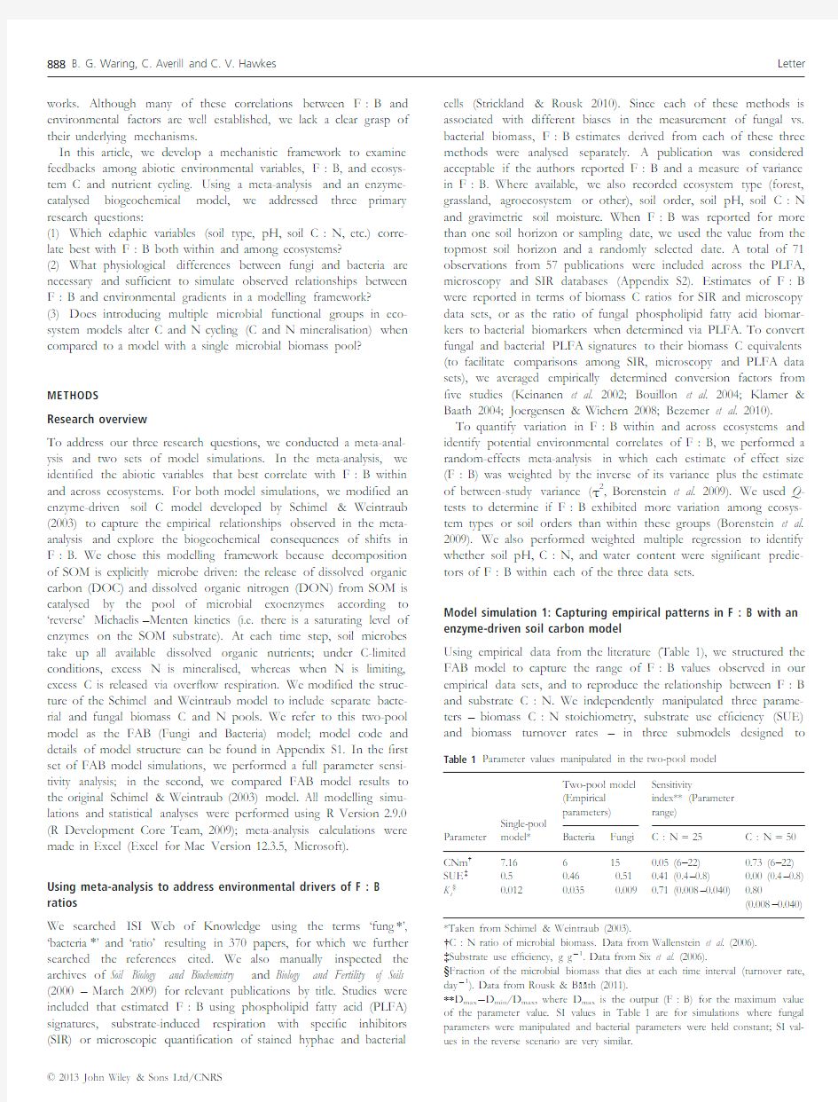
Using empirical data from the literature(Table1),we structured the FAB model to capture the range of F:B values observed in our empirical data sets,and to reproduce the relationship between F:B and substrate C:N.We independently manipulated three parame-ters–biomass C:N stoichiometry,substrate use ef?ciency(SUE) and biomass turnover rates–in three submodels designed to Table1Parameter values manipulated in the two-pool model
Parameter
Single-pool
model*
Two-pool model
(Empirical
parameters)
Sensitivity
index**(Parameter
range)
Bacteria Fungi C:N=25C:N=50 CNm?7.166150.05(6–22)0.73(6–22) SUE?0.50.460.510.41(0.4–0.8)0.00(0.4–0.8) K t§0.0120.0350.0090.71(0.008–0.040)0.80
(0.008–0.040) *Taken from Schimel&Weintraub(2003).
?C:N ratio of microbial biomass.Data from Wallenstein et al.(2006).
?Substrate use ef?ciency,g gà1.Data from Six et al.(2006).
§Fraction of the microbial biomass that dies at each time interval(turnover rate, dayà1).Data from Rousk&B a ath(2011).
**D maxàD min/D max,where D max is the output(F:B)for the maximum value of the parameter value.SI values in Table1are for simulations where fungal parameters were manipulated and bacterial parameters were held constant;SI val-ues in the reverse scenario are very similar.
?2013John Wiley&Sons Ltd/CNRS
888B.G.Waring,C.Averill and C.V.Hawkes Letter
capture different degrees of trait variation between fungi and bacte-ria.Next,to explore dynamics of F:B along environmental gradi-ents when biomass stoichiometry,SUE and biomass turnover all differ between the two groups(as is likely to occur in real soils),we manipulated all of these traits simultaneously.
In the stoichiometry-only submodel,the C:N of bacterial and fun-gal biomass was the only physiological difference between the two groups.We set bacterial biomass C:N to6and fungal biomass C:N to15(Wallenstein et al.2006).All subsequent model versions also included differences in fungal and bacterial biomass stoichiome-try,since there is very strong empirical evidence of higher mean bio-mass C:N in fungi than bacteria(Paul&Clark1996;Harris et al. 1997;Wallander et al.2003).However,since both bacteria and fungi can exhibit wide variation in biomass C:N ratios,we also explored the consequences of increasing the difference in fungal vs.bacterial biomass stoichiometry while keeping overall microbial biomass C:N constant.To examine the biogeochemical consequences of varying substrate use ef?ciency between groups,the SUE submodel addition-ally included differences between SUE of bacteria and fungi,or SUE b and SUE f.To capture differences in biomass turnover rates between the fungal and bacterial biomass,in the turnover submodel we varied the proportion of bacterial and fungal biomass that died in each time interval,or K tb and K tf.
To explore how F:B responded to trait variation in the three model scenarios,we calculated mean F:B(as the ratio of C in fun-gal biomass to bacterial biomass)after equilibration(2500model time steps)across a range of SOM C:N values.Each simulation began with an F:B of1:1.These simulations were repeated with a range of biomass C:N,SUE and K t values so that sensitivity of F:B to variation in each of these parameters could be quanti?ed with the sensitivity index(SI)of Hoffman&Gardner(1983).
After we explored qualitative model behaviour in response to inde-pendent manipulation of each parameter,we further assessed how well our model captured relationships between F:B and C:N observed in the meta-analysis data set with multiple simultaneous dif-ferences in fungal and bacterial physiology.We modelled F:B across a range of substrate C:N using values of SUE f,SUE b,K tf and K tb obtained from empirical studies of bacterial and fungal CUE(which is the same as the SUE term in our model)and biomass turnover rates (Table1).Fungal and bacterial CUE values were taken from data summarised by Six et al.(2006)and biomass turnover rates were cal-culated from soil-speci?c data reported in Rousk&B a ath(2011). Because the relationship between F:B and C:N changed along a gradient of substrate C:N in the FAB model,we used piecewise regression techniques to estimate breakpoints(i.e.the value(s)of the independent variable at which the slope of the relationship changes).The same analysis was performed with the meta-analysis data set to determine if a similar change in slope occurs along a gra-dient of soil C:N.For piecewise regression analyses,we used the segmented package in R(Muggeo2012).
Model simulation2:Consequences of shifts in F:B for C and N cycling
Most soil C-cycling models include a single microbial biomass pool, ignoring potential interactions among functional groups.To understand how including separate bacterial and fungal biomass pools might affect model predictions,we simulated respiration?uxes,inorganic N pools and total microbial biomass pools over a range of substrate C:N ratios using our stoichiometry-only soil C and N model.The resulting pool sizes were compared to those in the similarly structured original model(Schimel&Weintraub2003)with a single microbial biomass pool.For these simulations,we set the microbial biomass C:N parameter in the single-pool model to match the overall biomass C:N value predicted by the two-pool model after F:B had equilibrated. We calculated the size of fungal and bacterial C and N pools,enzyme C pools and the size of the inorganic N pool at t=2500(after the model had completely equilibrated),as well as cumulative respiration and cumulative over?ow respiration over the entire simulation. Because bacteria and fungi have different biomass stoichiometries, they may either compete for resources or exploit them in a comple-mentary fashion,depending on the C:N ratio of the substrate.There-fore,we also asked whether total microbial biomass production in simulations including both fungi and bacteria exceeded those including either fungal or bacterial groups alone along a gradient of resource C:N.This is analogous to overyielding effects in diversity–productiv-ity experiments(e.g.Hooper&Dukes2004).We computed mean microbial biomass C after2500daily time steps for bacteria only,fungi only and both groups,beginning with an F:B of1:1and repeated across a range of substrate C:N values.Simulation outcomes were used to calculate relative yield total(RYT)as an index of overyielding (Hooper&Dukes2003):RYT?
P s
i?1
RY i;where s is the number of species(or in this case,functional groups).Here,RY i=O i/M i,or mixture yield of functional group i divided by the monoculture yield of functional group i.Over-yielding is indicated when RYT>1,with production by two or more functional groups in mixture greater than expectations arising from production by each group alone.Here,the quantity[(RYTà1)9100]indicates the percentage increase or decrease in the microbial biomass C pool when fungi and bacteria co-exist,relative to fungal and bacterial monocultures.
RESULTS
Meta-analysis
Across studies,mean ratios of fungal biomass C(mg gà1)to bacte-rial biomass C were consistently>1.Mean F:B was7.50?4.42 (1SE,n=52)in the PLFA data set,1.12?1.10in the microscopy data set(n=13)and4.00?2.97in the SIR data set(n=6).In the PLFA data set,F:B varied across ecosystem types (Q=149.72,P<0.001),and was68%greater in forests than grass-lands(Table2).Agroecosystems had similar F:B on average com-pared to grasslands,but were more variable perhaps in part due to the smaller sample size(Table2).Similar patterns were observed in the microscopy(Q=31.66,P<0.001)and SIR data sets(Appen-dix S3,Tables S1and S2),although the latter had too few data points for analysis via Q-tests.In multiple regression analyses,the only signi?cant predictor of F:B was soil C:N,which explained 22%of the variance in F:B in the PLFA data set(Appendix S3, Fig.S1)and33%of the variance in the microscopy data set.
Capturing variation in F:B along a soil C:N gradient with an enzyme-driven soil carbon model
Qualitative model behaviour
In the stoichiometry-only submodel,when fungi and bacteria had equal SUE and K t,but different biomass C:N ratios,F:B only
?2013John Wiley&Sons Ltd/CNRS
Letter Modelling soil fungal–bacterial ratios889
varied between 1.0and 2.5regardless of substrate C :N.To achieve F:B consistently >1as observed in the meta-analysis data sets required either fungal SUE to be greater than bacterial SUE or fungal biomass turnover rates to be lower than bacterial turnover rates (Fig.1).When fungi had a higher biomass C :N or SUE,or a lower K t than bacteria,a positive relationship between F :B and substrate C :N was obtained (Fig.1),matching empirical data sets.At very high substrate C :N values (>45),F :B no longer increased with C :N,yielding a saturating relationship between F :B and substrate C :N over a broad range of substrate stoichi-ometries.There was no empirical evidence to support or invalidate this pattern in real soils;however,as the highest substrate C :N in the meta-analysis data set was 40.8.
The sensitivity of F :B to each physiological parameter depended on substrate C :N (Table 1).When both groups were limited by C (substrate C :N =25),the model was more sensitive to variation in K t than SUE or biomass C :N ratios.For example,a 50%decrease in fungal K tf vs.K tb yielded a 52.6%increase in F :B,whereas the same percentage change in SUE f vs.SUE b increased F :B by 39.4%.The fungal-to-bacterial ratio changed by less than 10%when fungal and bacterial biomass C :N were allowed to range from 6to 22and 3to 15respectively.In contrast,when both
groups were limited by N (substrate C :N =50),model results were sensitive to K t and biomass C :N ratios for each functional group,but not SUE (Table 1).Under conditions of N-limitation,the FAB model was not only sensitive to the absolute values of bio-mass C :N ratios but also to the degree of divergence between fungal and bacterial biomass stoichiometry.At a substrate C :N of 50,predicted F :B scaled linearly with the per cent difference between fungal and bacterial biomass C :N (Appendix S3,Fig.S2).Quantitative model behaviour
When parameterised with SUE and K t values estimated from the lit-erature,our model predicted that F :B should vary between 3.13and 9.72along a gradient of substrate C :N (Fig.2b).These values are within the range of F :B observed in the meta-analysis (Fig.2a).The FAB model predicted a signi?cant change in the slope of the relationship between F :B and C :N at a substrate C :N of 33.96?0.099(Davies Test,P <0.001).At lower sub-strate C :N values,the slope was estimated to be 0.014?0.002,whereas above a substrate C :N of 34,the slope was 0.580?0.010.After excluding a signi?cant outlier (Grubbs Test,P =0.001)in the meta-analysis data set,we also found a signi?cant breakpoint in the slope of the relationship at a soil C :N of 18.4(Davies Test,P =0.017).At lower soil C :N values,the slope was estimated to be à0.368?0.260(not signi?cantly different from 0),whereas at C :N values above the breakpoint,the slope was esti-mated to be 0.548?0.149.
Table 2Ratios of fungal to bacterial biomass C in the meta-analysis PLFA data
set Biome
Sample size F :B ?1SD*Range Forest 248.07?0.740.77–34.25Grassland
17 5.40?0.64 1.07–15.34Agroecosystem
8 5.25?1.780.61–11.68Other (Desert,Tundra)
3
1.23
?0.59
0.38–3.49
*Fungal :bacterial PLFA ratios have been converted to a fungal :bacterial bio-mass C ratio using a conversion factor of 27.4(Keinanen et al.2002;Bouillon et al.2004;Klamer &Baath 2004;Joergensen &Wichern 2008;Bezemer et al.
2010).
?2013John Wiley &Sons Ltd/CNRS
890B.G.Waring,C.Averill and C.V.Hawkes
Letter
Consequences of shifts in F:B for C and N cycling
Although over?ow respiration was substantially greater in the two-pool vs.the single-pool model at intermediate substrate C:N (Fig.3a),total respiration?uxes did not differ(Fig.3b).Inorganic and microbial biomass N pools diverged between the one-and two-pool models across a broad range of substrate C:N(Fig.3c and d).In the two-pool model,net mineralisation was generally lower because bacteria immobilised the inorganic N mineralised by fungi,but some net mineralisation was still predicted to occur at a substrate C:N of40.In contrast,the one-pool model predicted that all inorganic N would be immobilised by the microbial biomass by C:N of40.
When fungi and bacteria differed only in biomass stoichiometry, the RYT index was1where both fungi and bacteria were limited by the same nutrient,indicating that bacteria and fungi were compet-ing for available resources(Appendix S3,Table S3).Consequently, microbial biomass C was reduced by up to3%in mixtures compared to when bacteria and fungi were alone(Fig.4).In contrast,overyield-ing(RYT>1)occurred at a substrate C:N of35–40,when bacteria were limited by N and fungi by C,indicating facilitative interactions between the two groups that increased microbial biomass C as much as15%(Fig.4).Similar patterns were observed when SUE f>SUE b and when K tf DISCUSSION By integrating differences between fungal and bacterial stoichiome-try and turnover time in our modelling framework,we show that these physiological traits are suf?cient to qualitatively reproduce the relationship between soil C:N and F:B observed in our meta-analysis data set.Therefore,the FAB model demonstrates that mea-surable differences in fungal and bacterial physiology can drive the relative abundance of these two groups along environmental gradi-ents.Changes in F:B can also impact nutrient cycling at the eco-system scale by altering dynamics of over?ow respiration and net N mineralisation. ?2013John Wiley&Sons Ltd/CNRS Letter Modelling soil fungal–bacterial ratios891 Meta-analysis of environmental drivers of F:B Although fungi tended to dominate over bacteria in all data sets, the magnitude of F:B was sensitive to the analytical method used. Lower estimates were obtained via microscopy vs.PLFAs or SIR with selective inhibitors,likely because microscopic quanti?cation underestimates total fungal biomass(Joergensen&Wichern2008). However,alternate methods are subject to other problems:SIR can produce erroneous estimates of F:B if‘selective’inhibitors affect non-target organisms(Anderson&Domsch1973).Converting PLFA signatures to biomass C can be problematic due to an unknown degree of interspeci?c variation in fatty acid:biomass ratios,and varying extraction ef?ciencies of speci?c PLFAs among soil types(Joergensen&Wichern2008).Variation in PLFA conver-sion factors across ecosystems may contribute to the scatter in F:B values at low soil C:N ratios(Fig.2a).Although each ana-lytical method carries its own unique bias,all quanti?cation methods yielded similar relationships between F:B and environmental vari-ables. Within each data set,soil F:B was largely controlled by resource stoichiometry.The relationship between soil C:N and F:B in the meta-analysis supports empirical trends documented both within and across ecosystems(Lauber et al.2008;Fierer et al.2009).The change in the slope of the relationship between F:B and soil C:N is consistent with the idea of a threshold element ratio (Sterner&Elser2002;Allen&Gillooly2009)at which microbial metabolism switches from dependence upon energy(C)availability to nutrient availability.Because fungi and bacteria have different biomass stoichiometry,they are likely to exhibit different threshold element ratios.Thus,along a gradient of substrate C:N,relation-ships between F:B and soil C:N should change when both fungi and bacteria are C-limited,when both are N-limited,and when the two groups differ in element limitation. Effects of biomass stoichiometry,substrate use ef?ciency and biomass turnover rates on F:B ratios FAB model simulations suggest that differences in biomass stoichi-ometry have minimal in?uence on F:B when C is limiting,but set the upper bounds of the F:B ratio under conditions of nutrient limitation.Meanwhile,differences in fungal vs.bacterial SUE have a small effect on C-cycling when C is limiting,and do not control the magnitude of F:B when nutrients are limiting.In contrast,modify-ing fungal and bacterial biomass turnover rates(K t)to re?ect empir-ical data yields sensible F:B values across all C:N ratios.The fungal advantage conferred by K t is increased at high substrate C:N values that promote N-limitation,due to decreased losses of N from biomass turnover and recycling associated with a lower bio-mass turnover rate. The FAB model simulations suggest that in soils where one func-tional group is dominant,bacteria and fungi are likely to exhibit sys-tematic differences in biomass stoichiometry,substrate use ef?ciency,and/or turnover rates.Yet some empirical studies dem-onstrate that fungi and bacteria can exhibit very similar physiologies under some conditions.For instance,fungi and bacteria have identi-cal growth ef?ciencies when metabolising simple substrates(Thiet et al.2006),although interspeci?c differences in growth rate may become more pronounced on complex organic substrates(e.g. Goldfarb et al.2011).Fungal and bacterial biomass stoichiometry can also exhibit some degree of overlap(Strickland&Rousk2010). Identi?cation of the environmental or ecological conditions that drive divergence between bacterial and fungal physiology is outside the scope of our analysis(but see de Boer et al.2005).Nevertheless, because our model results are sensitive to the magnitude of the dif-ference between fungal and bacterial SUE,turnover time,and bio-mass C:N,we suggest that the degree of physiological trait overlap between these two groups can in?uence ecosystem C and N cycling. Comparing model results with empirical patterns Modelled differences in fungal and bacterial biomass stoichiometry and biomass turnover rates were suf?cient to explain empirical pat-terns in F:B along gradients of substrate C:N.Our model repro-duced a breakpoint in the relationship between F:B ratio and soil C:N that was also observed in the meta-analysis,demonstrating signi?cant differences in the slope of the relationship before and after a threshold soil C:N.Although the slopes of the empirical and modelled relationships between F:B and substrate C:N are the same,the location of this threshold differed in the model vs.in the empirical data set,potentially re?ecting differences in substrate use ef?ciencies or fungal and bacterial biomass stoichiometry in the model vs.the studies cited in the meta-analysis.In addition,the FAB model failed to simulate the extremely high F:B values (F:B>10)observed in our meta-analysis data set.Although these high values may be artefacts of inaccurate PLFA:biomass conver-sion factors,such pronounced fungal dominance may also re?ect other potential drivers of F:B.For instance,F:B may be increased by high plant investment in mycorrhizal fungi in low-fer-tility systems,or by inputs of highly recalcitrant litter rich in lignin, which only fungi can degrade(de Boer et al.2005). Our meta-analysis and model simulations provide insight into the idiosyncratic responses of F:B to manipulation of soil C:N in the?eld and laboratory.In response to short-term fertilisation with inorganic N,F:B either decreases or does not change(Strickland &Rousk2010).FAB model results provide a potential mechanism for these variable responses:shifts in F:B following changes in soil C:N are likely dependent upon initial resource stoichiometry and the location of the threshold element ratio.When both fungi and bacteria are C-limited,altering soil C:N will not in?uence their relative abundance.However,when both groups are N-limited, decreasing the availability of N relative to C should enhance fungal dominance and vice versa.Therefore,responses of F:B to fertilisa-tion with C or N may not be fully predictable unless the nutrient limitation status of each functional group is known. Our two-pool model captured competitive and facilitative interac-tions among functional groups that in?uence the size of the total microbial biomass.When fungi are C-limited but bacteria are N-lim-ited,the model predicts that N mineralisation by fungi fuels bacte-rial growth,allowing for overyielding to occur.In accordance with this model prediction,laboratory experiments on decomposing leaf litter show that bacterial populations grow faster in the presence of fungi,while fungi grow more rapidly in isolation(Roman ?et al. 2006).Furthermore,bacterial inhibition of fungal growth is stronger on low C:N substrates(Rousk&B a ath2007;Rousk et al.2008), consistent with the model prediction that competition between bac-teria and fungi should be strongest at very low or very high sub-strate C:N.Finally,as F:B changed in the FAB model,so did ?2013John Wiley&Sons Ltd/CNRS 892B.G.Waring,C.Averill and C.V.Hawkes Letter the emergent C:N ratio of the microbial biomass,consistent with empirical observations.However,an increase in the C:N ratio of the microbial biomass does not correspond with greater rates of N mineralisation,as it does in the original Schimel&Weintraub (2003)model.When bacteria and fungi are limited by different ele-ments,bacteria continue to immobilise inorganic N mineralised by fungi,resulting in lower mineralisation rates even as total microbial biomass C:N increases.This is consistent with an empirically determined negative correlation between F:B and gross mineralisa-tion rates across forest types(H€o gberg et al.2007)and is a qualita-tive departure from current biogeochemical models with a single microbial functional group.To obtain this pattern in a model simu-lation,community structure must be explicit in the model. Biogeochemical insights from the FAB model Differences in fungal and bacterial physiology may have important consequences for ecosystem-level C and N cycling.The largest bio-geochemical difference between our modi?ed FAB model and the original Schimel&Weintraub(2003)model is observed in N miner-alisation rates.When one functional group is N-limited while the other is C-limited,the community N immobilisation rate is effec-tively increased as bacteria immobilise the fungal mineralisation product.N mineralisation rates are tightly coupled with NPP both empirically(Reich et al.1997)and in ecosystem models(Melillo& Gosz1983),suggesting that including differences in fungal and bac-terial physiology in a full ecosystem model would substantially in?u-ence NPP simulations.Additionally,our model demonstrates competitive and facilitative interactions between fungi and bacteria can alter the size of the total microbial biomass,with clear conse-quences for the cycling of C,N and other nutrients through soils. Our community-explicit FAB model does not predict large differ-ences in total respiration?uxes relative to those reported in the ori-ginal Schimel&Weintraub(2003)model.Changes in the magnitude of over?ow respiration at intermediate substrate C:N do not sub-stantially alter total respiratory?uxes,since increases in over?ow respiration are offset by decreases in maintenance respiration.How-ever,it is not clear that all C in excess of microbial demand is min-eralised as CO2,as our model assumes.In at least some bacterial taxa,C uptake in excess of demand can be released as simple organic C compounds rather than as CO2(e.g.Russell&Cook 1995;Xu et al.1999).Therefore,depending upon the ultimate fate of excess C uptake in soil,differences in over?ow metabolism between the one-and two-pool models may have consequences for soil C storage. CONCLUSIONS Although soil microbial communities are characterised by an over-whelming variety of bacterial and fungal taxa,the FAB model dem-onstrates that this diversity can be consolidated into broad yet meaningful and quanti?able functional groups whose relative abun-dance impacts ecosystem processes.The enzyme-driven FAB model was able to capture empirical relationships between F:B and soil C:N across ecosystems,highlighting mechanistic links between microbial physiology,resource availability,and F:B.By integrating multiple physiological differences between fungi and bacteria in FAB model,we explain how seemingly inconsistent responses of F:B to N fertilisation may actually be consistent within a broader theoretical framework.Furthermore,incorporating multiple func-tional groups in our biogeochemical model allows for both competi-tive and facilitative interactions to develop within the community, slowing the net mineralisation rate and enhancing over?ow respira-tion under some conditions.Because of the tight connection between net mineralisation and NPP(Reich et al.1997),we argue that representing bacteria and fungi explicitly may be important for simulating responses of NPP and therefore ecosystem C balance to environmental change in large-scale biogeochemical models. ACKNOWLEDGEMENTS The authors thank Adrien Finzi,Heather McGray and three anon-ymous referees for thoughtful comments that substantially improved the manuscript.The authors would like to extend many thanks to the researchers who generously provided raw data for the meta-analysis:Anne Kasurinen,Annemieke van der Wal,Asa Frostegard,Beligh Mechri,Bob Larkin,Bryan Grif?ths,Chantal Hamel,Chih-Yu Chiu,Dagmar Tscherko,David Lipson,David Myrold,Diana Ebersberger,Donald Klein,Erica Smithwick,Fang-Hao Wan,Franciska deVries,Greet Ruysschaert.Hannu Fritze, Heidi Anderson,Henk Velvis,Hongbang Niu,Huaiying Yao,Ilya Yevdokimov,Irene Unger,James Entry,Jan Hassink,Jeff Smith, Joeke Postma,Joern Alphei,Johannes Rousk,K Sakamoto,Katari-na Hedlund,Kirsten Kuesel,Kris Brye,Liliane Ruess,Mark Wil-liams,Martin Romantschuk,Mary Stromberger,Matt Busse,Matt Wallenstein,Michael Mullen,Mona Hogsbert,Nadezhda Ananyeva, Nick van Eekeren,Paivi Merila,Petra Marschner,Rebecca Dre-novsky,Richard Bardgett,Richard Dick,Robert Bjork,Robert Bradley,Roger Burke,Sara Elfstrand,Sarah Kemmit,Sharon Bill-ings,Sherri Morris,Stefan Dekker,Stephanie Yarwood,Susyan Evgeny,Taina Pennanen,Ute Hamer,Valentyna Krashevska,Va-nessa Bailey,Wes Wood and Yosef Steinberger.This study is based on work supported by the National Science Foundation Graduate Research Fellowship Programme(Grant DGE-1110007 to BGW and CA).Any opinions,?ndings and conclusions or rec-ommendations expressed in this material are those of the authors and do not necessarily re?ect the views of the National Science Foundation. AUTHORSHIP BGW and CA conceived the conceptual framework,wrote the model,and interpreted model simulation results.BGW and CVH performed the meta-analysis.BGW and CA wrote the?rst draft of the manuscript;all authors contributed to revisions.The authors declare no con?icts of interest. REFERENCES Allen, A.&Gillooly,J.(2009).Towards an integration of ecological stoichiometry and the metabolic theory of ecology to better understand nutrient cycling.Ecol.Lett.,12(5),369–384. Allison,S.(2012).A trait-based approach for modeling microbial litter decomposition.Ecol.Lett.,15,1058–1070. Anderson,J.P.E.&Domsch,K.H.(1973).Quanti?cation of bacterial and fungal contributions to soil respiration.Archiv.fur Mikrobiologie,93,113–127. B a ath,E.&Anderson,T.(2003).Comparison of soil fungal/bacterial ratios in a pH gradient using physiological and PLFA-based techniques.Soil Biol.Biochem., 35,955–963. ?2013John Wiley&Sons Ltd/CNRS Letter Modelling soil fungal–bacterial ratios893 Bailey,V.,Smith,J.&Bolton,H.(2002).Fungal-to-bacterial ratios in soils investigated for enhanced C sequestration.Soil Biol.Biochem.,34,997–1007. Bezemer,T.M.,Fountain,M.T.,Barea,J.M.,Christensen,S.,Dekker,S.C.,Duyts, H.et al.(2010).Divergent composition but similar function of soil food webs of individual plants:plant species and community effects.Ecology,91,3027–3036. de Boer,W.,Folman,L.,Summerbell,R.&Boddy,L.(2005).Living in a fungal world:impact of fungi on soil bacterial niche development.FEMS Microbiol. Rev.,29,795–811. Borenstein,M.,Hedges,L.V.,Higgins,J.P.T.&Rothstein,H.R.(2009). Introduction to Meta-Analysis.Wiley,West Sussex. Bouillon,S.,Moens,T.,Koedam,N.,Dahdouh-Guebas, F.,Baeyens,W.& Dehairs,F.(2004).Variability in the origin of carbon substrates for bacterial communities in mangrove sediments.FEMS Microbiol.Ecol.,49,171–179. Fierer,N.,Strickland,M.,Liptzin,D.,Bradford,M.&Cleveland,C.(2009). Global patterns in belowground communities.Ecol.Lett.,12,1238–1249. Frey,S.D.,Elliott,E.T.&Paustian,K.(1999).Bacterial and fungal abundance and biomass in conventional and no-tillage agroecosystems along two climatic gradients.Soil Biol.Biochem.,31,573–585. Frey,S.,Knorr,M.,Parrent,J.&Simpson,R.(2004).Chronic nitrogen enrichment affects the structure and function of the soil microbial community in temperate hardwood and pine forests.For.Ecol.Manage.,196,159–171. Goldfarb,K.C.,Karaoz,U.,Hanson, C.A.,Santee, C.A.,Bradord,M.A., Treseder,K.K.et al.(2011).Differing growth responses of soil bacterial taxa to carbon substrates of varying chemical recalcitrance.Front.Microbiol.,2, 1–10. Harris, D.,Voroney,R.P.&Paul, E.A.(1997).Measurement of microbial biomass N:C by chloroform fumigation-incubation.Can.J.Soil Sci.,77,507–514. Hoffman, F.O.&Gardner,R.H.(1983).Evaluation of uncertainties in environmental radiological assessment models.In:Radiological Assessments:A Textbook on Environmental Dose Assessment(eds Till,J.E.&Meyer,H.R.).U.S. Nuclear Regulatory Commission,Washington,DC,pp.11-1–11-55. H€o gberg,M.,Chen,Y.&H€o gberg,P.(2007).Gross nitrogen mineralisation and fungi-to-bacteria ratios are negatively correlated in boreal forests.Biol.Fertil. Soils,44,363–366. Hooper,D.&Dukes,J.(2004).Overyielding among plant functional groups in a long-term experiment.Ecol.Lett.,7,95–105. Joergensen,R.&Wichern, F.(2008).Quantitative assessment of the fungal contribution to microbial tissue in soil.Soil Biol.Biochem.,40,2977–2991. Keinanen,M.M.,Korhonen,L.K.,Lehtola,M.J.,Miettinen,I.T.,Martikainen, P.J.,Vartiainen,T.et al.(2002).The microbial community structure of drinking water bio?lms can be affected by phosphorus availability.Appl. Environ.Microbiol.,68,434–439. Klamer,M.&Baath,E.(2004).Estimation of conversion factors for fungal biomass determination in compost using ergosterol and PLFA18:2w6,9.Soil Biol.Biochem.,36,57–65. Lauber,C.,Strickland,M.,Bradford,M.&Fierer,N.(2008).The in?uence of soil properties on the structure of bacterial and fungal communities across land-use types.Soil Biol.Biochem.,40,2407–2415. McGill,W.B.,Hunt,H.W.,Woodmansee,R.G.&Ruess,J.O.(1981). PHOENIX:a model of the dynamics of carbon and nitrogen in grassland soils.In:Terrestrial Nitrogen Cycles:Processes,Ecosystem Strategies,and Management Impacts,Ecol.Bull.33(edsClark, F.E.&Rosswall,T.).Swedish Natural Science Research Council,Stockholm,Swedenpp.49–115. McGuire,K.L.&Treseder,K.K.(2010).Microbial communities and their relevance for ecosystem models:decomposition as a case study.Soil Biol. Biochem.,42,529–535. Melillo,J.M.&Gosz,J.R.(1983).Interactions of biogeochemical cycles in forest ecosystems.In:The Major Biogeochemical Cycles and Their Interactions(eds Bolin,B. &Cook,R.B.).John Wiley and Sons,New Yorkpp.177–222. Moorhead,D.&Sinsabaugh,R.L.(2006).A theoretical model of litter decay and microbial interaction.Ecol.Monogr.,76,151–174.Muggeo,V.M.R.(2012).Segmented:Segmented relationships in regression models with breakpoints/changepoints estimation. Orwin,K.H.,Kirschbaum,M.U.F.,St John,M.G.&Dickie,I.A.(2011).Organic nutrient uptake by mycorrhizal fungi enhances ecosystem carbon storage:a model-based assessment.Ecol.Lett.,14,493–502. Paul,E.A.&Clark,F.E.(1996).Soil Microbiology and Biochemistry.Academic Press, San Diego. R Development Core Team(2009).R:A language and environment for statistical computing.R Foundation for Statistical Computing,Vienna,Austria. Reich,P.B.,Grigal, D.F.,Aber,J.D.&Gower,S.T.(1997).Nitrogen mineralization and productivity in50hardwood and conifer stands on diverse soils.Ecology,78,335–347. Roman ?,A.,Fischer,H.,Mille-Lindblom,C.&Tranvik,L.(2006).Interactions of bacteria and fungi on decomposing litter:differential extracellular enzyme activities.Ecology,87,2559–2569. Rousk,J.&B a ath,E.(2007).Fungal and bacterial growth in soil with plant materials of different C/N ratios.FEMS Microbiol.Ecol.,62,258–267. Rousk,J.&B a ath,E.(2011).Growth of saprotrophic fungi and bacteria in soil. FEMS Microbiol.Ecol.,78,17–30. Rousk,J.,Demoling,L.,Bahr,A.&B a ath,E.(2008).Examining the fungal and bacterial niche overlap using selective inhibitors in soil.FEMS Microbiol.Ecol., 63,350–358. Russell,J.B.&Cook,G.M.(1995).Energetics of bacterial growth:balance of anabolic and catabolic reactions.Microbiol.Rev.,59,48–62. Schimel,J.&Weintraub,M.(2003).The implications of exoenzyme activity on microbial carbon and nitrogen limitation in soil:a theoretical model.Soil Biol. Biochem.,35,549–563. Schlesinger,W.H.(1997).Biogeochemistry:An Analysis of Global Change.Academic Press,San Diego. Six,J.,Frey,S.D.,Thiet,R.K.&Batten,K.M.(2006).Bacterial and fungal contributions to carbon sequestration in agroecosystems.Soil Sci.Soc.Am.J., 70,555–569. Sterner,R.W.&Elser,J.J.(2002).Ecological Stoichiometry:The Biology of Elements from Molecules to the Atmosphere.Princeton University Press,Princeton. Strickland,M.&Rousk,J.(2010).Considering fungal:bacterial dominance in soils-Methods controls and ecosystem implications.Soil Biol.Biochem.,4,1385–1395. Swift,M.J.,Heal,O.W.&Anderson,J.M.(1979).Decomposition in Terrestrial Ecosystems.Blackwell,Oxford. Thiet,R.,Frey,S.&Six,J.(2006).Do growth yield ef?ciencies differ between soil microbial communities differing in fungal:bacterial ratios?Reality check and methodological issues.Soil Biol.Biochem.,38,837–844. Wallander,H.,Nilsson,L.O.,Hagerberg,D.&Rosengren,U.(2003).Direct estimates of C:N ratios of ectomycorrhizal mycelia collected from Norway spruce forest soils.Soil Biol.Biochem.,35,997–999. Wallenstein,M.D.,McNulty,S.,Fernandez,I.J.,Boggs,J.&Schlesinger,W.H. (2006).Nitrogen fertilization decreases forest soil fungal and bacterial biomass in three long-term experiments.For.Ecol.Manage.,222,459–468. Xu,B.,Jahic,M.&Enfors,S.(1999).Modeling of over?ow metabolism in batch and fed-batch cultures of Escherichia coli.Biotechnol.Prog.,15,81–90. SUPPORTING INFORMATION Additional Supporting Information may be downloaded via the online version of this article at Wiley Online Library(https://www.doczj.com/doc/9f5799097.html,). Editor,Marcel Holyoak Manuscript received24December2012 First decision made1February2013 Second decision made2April2013 Manuscript accepted15April2013 ?2013John Wiley&Sons Ltd/CNRS 894B.G.Waring,C.Averill and C.V.Hawkes Letter 微生物英文名词解释 1.Mycoplasma:The mycoplasma are a group of the smallest organisms without cell wall that can be free-living in nature, can pass through bacterial filter and also grow on laboratory media. 2.Chlamydia:Chlamydia are small Gram-negative bacteria which are obligate intercellular parasites like virus, but differ from them in that they have both RNA and DNA, ribosome, cell wall, and divided by binary fission. 3.L forms of bacteria:In osmotically protective media, removal of the bacterial wall with lysozyme or penicillin liberate protoplasts from Gram-positive cells and spheroplasts from Gram-negative cells. If such wall-defective cells are able to grow and divide, they are called L forms. L forms are difficult to cultivate. They require a special media. Some L form can revert tothe normal bacillary form. L form in the host may produce chronic infection that are relatively resistant to antibiotic treatment. 4.Capsule:Many bacteria synthesize large amounts of extracellular polymer when growing in their natural environments. When the polymer forms a condensed, well-defined layer closely surrounding the cell, it is called the capsule. With one known exception (the polypeptide capsule), the polymer is polysaccharide. 5.Pyrogen:This is a fever-producing substance synthesized by bacteria. In fact, it is the lipopolysaccharide of Gram-negative bacteria. For the injectable medicament, it is especially important to avoid the contamination of pyrogen in the course of pharmic production. 6.Exotoxin:Exotoxins are proteins produced inside Gram-positive bacteria cells and secreted into the environment. These toxins are some of the strongest poisons known to man and cause violent reactions in host organisms. 7.Endotoxin:Endotoxins are made up of lipids and carbohydrates associated with the outer membrane of gram-negative bacteria. These toxins usually produce fever, weakness, and capillary damage. 8.Disinfection: Reduce or eliminate pathogens病原体in or on inanimate无生命 的objects to a safe level, which are no longer health hazard危险. 9.Sterilization: A physical or chemical process that completely destroys or removes all microbial life,including bacteria spore and viruses. 10.Antisepsis: Use chemical agents to inhibit or destroy the growth of microorganisms on skin or other living tissue. 11.Plasmids:Plasmids are small genetic elements that replicate independently of the bacterial chromosome. Most plasmids are circular, double-stranded DNA molecules varying from 1,500 to 400,000 base pairs. Like the bacterial chromosomal DNA, they can autonomously replicate and as such are referred to as replicons. 12.Transformation:It is the process by which bacteria take up fragments of naked DNA and incorporate them into their genomes. During transformation, DNA 常见微生物 界(Domain)、门(Phylum)、纲(Class)、目(Order)、科(Family)、属(Genus)、种(Species)中英文对照 界(Domain)Bacteria细菌 Archaea古生菌 门(Phylum)Proteobacteria 变形菌门 Bacteroidetes 拟杆菌门 Actinobacteria 放线菌门Gemmatimonadetes 芽单胞菌门Acidobacteria 酸杆菌门Planctomycetes 浮霉菌门Verrucomicrobia 疣微菌门 Chloroflexi 绿弯菌门 Nitrospirae 硝化螺旋菌门 Firmicutes 厚壁菌门 Chlorobi 绿菌门 Cyanobacteria 蓝藻细菌门Fibrobacteres 纤维杆菌门 Elusimicrobia 迷踪菌门Armatimonadetes 装甲菌门Euryarchaeota 广古菌门 Chlamydiae 衣原体 Crenarchaeota 泉古菌门 Tenericutes 无壁菌门 Spirochaetes 螺旋体属 Alphaproteobacteria 甲型(α)变形杆菌纲Gammaproteobacteria 丙型变形菌纲Betaproteobacteria β-变形菌纲Actinobacteria 放线菌门、纲Cytophagia 纤维粘网菌Gemmatimonadetes 芽单胞菌门、纲Deltaproteobacteria δ-变形菌纲Acidobacteria-6 酸杆菌门Acidimicrobiia 酸微菌纲Verrucomicrobiae 疣微菌纲 Opitutae 丰佑菌纲 Nitrospira 消化螺菌属Thermomicrobia 热微菌门 Bacteroidia 拟杆菌纲 Bacilli 杆菌 Chloroflexi 绿弯菌门Anaerolineae 厌氧绳菌纲 Clostridia 梭状芽胞杆菌Elusimicrobia 迷踪菌门Ktedonobacteria 纤线杆菌纲Thermoplasmata 热原体纲Chlamydiia 衣原体Thaumarchaeota 奇古菌门 Mollicutes 柔膜菌纲Methanomicrobia 甲烷微菌纲Holophagae 全噬菌纲Spirochaetes 螺旋体属 Abdominal pain and Tenderness 14/03/02 STEVENSIU Learning Objectives 1 Describe the anatomy and physiology of the peritoneum, including the innervations of the visceral and parietal layers The peritoneum is a continuous, transparent serous membrane that lines the abdominopelvic cavity and invests the viscera. It consists of two continuous layers. 1.Parietal peritoneum- lining the internal surface of the abdominopelvic wall. 2.Visceral peritoneum – investing the viscera Visceral peritoneum supplied small type C pain fibers and can only response to chronic-aching suffering type of pain. These fibers can response to ischaemia, chemical stimuli, and spasm of a hollow viscus and over distention of a hollow viscus Parietal peritoneum is supplied by the spinal nerves not sympathetic nerves→ sharp localized somatic pain Learning objectives 2 Discuss conditions predisposing to spontaneous bacterial peritonitis (SBP) SBP= The inflammation of the peritoneum without a clear event such as bowel perforation that would account for the entry of the pathogenic organism Liver Cirrhosis is the main predisposing factor. Other conditions leading to ascites e.g. Fulminant hepatic failure, Congestive heart failure, liver metastases can also predispose to SBP. Pathogenesis 1.Seeding→ The exact pathogenic mechanism is unknown. Bacterial seeding is believed to involve haematogenous spread of organisms in a patient in whom diseased liver and altered portal circulation result in a defect in the usual filtration. 2.Growth--> The reduced levels of complement cascade and reduced opsonic and phagocytic properties of neutrophils in advanced liver cirrhosis in an excellent culture medium provided by the ascitic fluids, promote the growth of the organisms. Symptoms 1.Fever, Hypotension, Decreased or absent bowel sounds, Abdominal Pain and Abrupt onset of hepatic encephlopathy in patient with ascites Report 检验报告 OUR REF NO: ATS/MED/77/04/pl DATE: 31 March, 2004 Page 1 of 2 COMPANY: Okada Ecotech Pte Ltd Blk 1 Pasir Panjang Road Distripark #07-15/17 Alexandra 118478 Singapore 文件编号:ATS/MED/77/04/pl 日期: 2004年3月31日第1/2页 送检公司:冈田生态技术私人有限公司 地址:新加坡亚历山大分销园长滩路1区07-15/17号 邮编: 118478 DATE SAMPLE RECEIVED: 23 March, 2004 DATE ANALYZED: 23 March, 20004 DATE TEST COMPLETED: 31 March, 2004 SAMPLE DESCRIPTION: One bottle said to be Biovectrol 20EM (Indoor) was received with references :- 2004030801 BN 送检日期: 2004年3月23日 检验日期: 2004年3月23日 完成日期: 2004年3月31日 样品描述:名称为Biovectrol 20EM(室内用)样品一瓶,样品号: BN 2004030801 Method: Chemical Disinfectants & antiseptics – Basic Bactericidal Activity (BS EN 1040:1997) 检验方法:化学消毒剂和杀菌剂 – 基础试验(BS EN 1040:1997) Procedure: 检验程序: 1. 1ml of sample was transferred to 79ml of sterile water to make dilution of 1:79. Review Bacterial cold-shock proteins D.N.Ermolenko a,b and G.I.Makhatadze a,* a Department of Biochemistry and Molecular Biology, Penn State University College of Medicine, Hershey, Pennsylvania 17033 (USA), Fax: +17175317072, e-mail: makhatadze@https://www.doczj.com/doc/9f5799097.html, b A. N. Bach Institute of Biochemistry, Russian Academy of Sciences, Leninsky Prospect 33, 117071 Moscow (Russia) Received 26 April 2002; received after revision 27 May 2002; accepted 30 May 2002 Abstract.Members of a family of small cold-shock pro-teins (CSPs) are induced during bacterial cell response to a temperature decrease. Here we review available data about the structure, molecular properties, mechanism of induction and possible functions of CSPs. CSPs prefer-entially bind single-stranded RNA and DNA and appear to play an important role in cell physiology under both nor- CMLS, Cell. Mol. Life Sci. 59 (2002) 1902–19131420-682X/02/111902-12 ? Birkh?user Verlag, Basel, 2002 mal and cold-shock conditions. Although the function of CSPs in cold-shock adaptation has not yet been elucidated in detail, a number of experimental evidences suggests that CSPs bind messenger RNA (mRNA) and regulate ribosomal translation, rate of mRNA degradation and ter-mination of transcription. Introduction Bacteria respond to a decrease in temperature in a specific manner. A temperature downshift results in inhibition of cell growth and proliferation, and changes in protein ex-pression patterns [1]. The synthesis of most cellular pro-teins is inhibited after a decrease in temperature [1]. How-ever, a number of proteins are found to be induced under cold-shock conditions (for review see [2–5]). Expression of the cold-shock proteins reaches a maximum level dur-ing the phase of the cold-shock adaptation, the so-called acclimation. After this, synthesis of cold-shock proteins declines and a new steady-state level of protein expression is established which is lower than before the cold shock. Among cold-shock proteins, small homologous proteins [65–70 amino acid residues long) were found to be dom-inant under cold-shock conditions in different species of bacteria [6]. They are the most common cold-inducible proteins identified to date. The first protein described as a major protein induced upon decrease in temperature was CspA of Escherichia coli [7]. CspA production reached concentrations of 100 m M within 1–1.5h after the tem-perature downshift [7, 8]. Eight more proteins named in alphabetical order from CspB to CspI that are homologous to CspA are identified in E. coli [6]. CspB, CspG and CspI were found to be inducible by a temperature downshift similarly to CspA [9–11]. CspC and CspE are expressed at both high and low temperature [12], while CspD is in-duced during stationary phase and upon nutrition starva-tion [13]. In Bacillus subtilis three proteins homologous to E. coli CspA (CspB, CspC and CspD) are induced upon temperature downshift [14, 15]. CSP-like proteins were found in more than 50 other bacterial species [2, 16]. The first eukaryotic true homologue of CSPs, protein Cla h 8,was recently identified in the mould Cladosporium herbarum [17]. It appears that proteins of the CspA fam-ily not only play a major role during cold-shock adaptation but also are important under normal growth conditions.Despite the fact that CSPs were extensively studied dur-ing the last decade, their exact function both at normal and cold-shock conditions is not elucidated yet. Below we will focus on the known molecular properties, mechanisms of Key words.ssDNA/RNA binding protein; RNA chaperone; regulation of transcription; regulation of translation; bac- terial cold shock; cold adaptation. EUROPEAN PHARMACOPOEIA 5.0 2.6.14.Bacterial endotoxins Maize starch 1.0g Sodium chloride 5.0g Agar,according to gelling power 10.0g to 15.0g Purified water 1000ml Hydrate the agar,dissolve by heating to boiling with continuous stirring.If necessary,adjust the pH so that after sterilisation it is 7.3±0.2.Sterilise by heating in an autoclave at 121°C for 15min.Allow to cool to 45-50°C;add,where necessary,gentamicin sulphate corresponding to 20mg of gentamicin base and pour into Petri dishes.Medium R (Lactose monohydrate sulphite medium) Pancreatic digest of casein 5.0g Yeast extract 2.5g Sodium chloride 2.5g Lactose monohydrate 10.0g Cysteine hydrochloride 0.3g Purified water 1000ml Dissolve,adjust to pH 7.1±0.1and fill to 8ml in 16mm ×160mm tubes containing a small Durham tube. Sterilise by heating in an autoclave at 121°C for 15min and store at 4°C. Before use,heat the medium for 5min in a water-bath and cool.Add to each tube 0.5ml of a 12g/l solution of sodium metabisulphite R and 0.5ml of a 10g/l solution of ferric ammonium citrate,both solutions being freshly prepared and filtered through membranes (pore size:0.45μm). Agar medium S (R2A)Yeast extract 0.5g Proteose peptone 0.5g Casein hydrolysate 0.5g Glucose 0.5g Starch 0.5g Dipotassium hydrogen phosphate 0.3g Magnesium sulphate,anhydrous 0.024g Sodium pyruvate 0.3g Agar 15.0g Purified water 1000ml Adjust the pH so that after sterilisation it is 7.2±0.2.Sterilise by heating in an autoclave at 121°C for 15min.NEUTRALISING AGENTS Neutralising agents may be used to neutralise the activity of antimicrobial agents.They may be added to buffered sodium chloride-peptone solution pH 7.0,preferably before sterilisation.If utilised their efficacy and non-toxicity towards micro-organisms are demonstrated. A typical neutralising fluid has the following composition: Polysorbate 8030g Lecithin (egg)3g Histidine hydrochloride 1g Peptone (meat or casein)1g Sodium chloride 4.3g Potassium dihydrogen phosphate 3.6g Disodium hydrogen phosphate dihydrate 7.2g Purified water 1000ml Sterilise by heating in an autoclave at 121°C for 15min.If the solution has insufficient neutralising capacity the concentration of polysorbate 80or lecithin may be increased.Alternatively,the neutralisers mentioned in Table 2.6.13.-3may be added. Table 2.6.13.-3.–Inactivators for antimicrobial agents to be added to buffered sodium chloride-peptone solution pH 7.0Type of an-timicrobial agent Inactivator Concentration Comment Sodium laurilsulfate 4g/l Polysorbate 80and lecithin 30g/l and 3g/l Phenolics Egg yolk 5ml/l -50ml/l Add after sterilisation of buffered sodium chloride-peptone solution pH 7.0 Organo-mercurals Sodium thioglycolate 0.5g/l -5g/l Halogens Sodium thiosulphate 5g/l Quaternary ammonium compounds Egg yolk 5ml/l -50ml/l Add after sterilisation of buffered sodium chloride-peptone solution pH 7.001/2005:206142.6.14.BACTERIAL ENDOTOXINS The test for bacterial endotoxins is used to detect or quantify endotoxins of gram-negative bacterial origin using amoebocyte lysate from horseshoe crab (Limulus polyphemus or Tachypleus tridentatus ).There are 3techniques for this test:the gel-clot technique,which is based on gel formation;the turbidimetric technique,based on the development of turbidity after cleavage of an endogenous substrate;and the chromogenic technique,based on the development of colour after cleavage of a synthetic peptide-chromogen complex. The following 6methods are described in the present chapter:Method A.Gel-clot method:limit test Method B.Gel-clot method:semi-quantitative test Method C.Turbidimetric kinetic method Method D.Chromogenic kinetic method Method E.Chromogenic end-point method Method F.Turbidimetric end-point method Proceed by any of the 6methods for the test.In the event of doubt or dispute,the final decision is made based upon method A unless otherwise indicated in the monograph.The test is carried out in a manner that avoids endotoxin contamination.Apparatus Depyrogenate all glassware and other heat-stable apparatus in a hot-air oven using a validated process.A commonly used minimum time and temperature is 30minutes at 250°C.If employing plastic apparatus,such as microtitre plates and pipette tips for automatic pipetters,use apparatus shown to be free of detectable endotoxin and of interfering effects for the test. NOTE:In this chapter,the term ‘tube’includes all types of receptacles,for example microtitre plate wells . 綠色螢光蛋白質粒轉化實驗pGLO Bacterial Transformation 一.課前思考 骨骼是怎麼變疏鬆的 報章來源: https://www.doczj.com/doc/9f5799097.html,/shkj/2013-08/19/c_114413781.htm 報章日期: 2013年8月19日 日本大阪大學研究人員利用熒光蛋白標識實驗鼠的破骨細胞,首次“目睹”了破骨細胞破壞骨骼的情形,研究還發現破骨細胞在患骨質疏鬆症的實驗鼠體內與健康實驗鼠體內的明顯差異。這一發現將有助於研發出治療骨質疏鬆症和風濕病的新藥物。 破骨細胞具有促進骨骼代謝的作用。機體中存在著分解骨質的破骨細胞和形成骨骼的成骨細胞,正常情況下這兩種細胞的作用保持平衡。骨質疏鬆症患者體內這種平衡被打破,導致“破壞”快於“再造”。 大阪大學教授石井優率領的研究小組首次成功觀察到活體實驗鼠體內破骨細胞的破骨情形。研究發現,破骨細胞會以兩種狀態存在,一種是在骨組織表面釋放強酸溶解骨骼,被稱為R型;一種僅在骨組織表面移動但不破壞骨組織,被稱為N型。破骨細胞會在這兩種狀態之間轉換。 研究發現,在健康實驗鼠體內,R型破骨細胞佔總量的約40%,而在骨質疏鬆症實驗鼠體內,這一比例則上升到90%以上。如果使用常用治療藥物,雖然可以大幅降低破骨細胞的總數,但破骨細胞過少會導致骨質脆弱。 思考: 1.在研究人體內部的細胞變化、轉移時可以利用什麼來觀察得知? _________________________________________________________________________________________ _________________________________________________________________________________________ 2.根據上述報導,試分析科學家運用了什麼技術去觀察骨骼細胞變化? _________________________________________________________________________________________ _________________________________________________________________________________________ 3.除了觀察骨骼細胞變化外,利用上述技術還可以做作怎樣的研究? _________________________________________________________________________________________ _________________________________________________________________________________________ 常见微生物的界、门、纲、目、科、属、种中英文对照 常见微生物 界(Domain)、门(Phylum)、纲(Class)、目(Order)、科(Family)、属(Genus)、种(Species)中英文对照 界(Domain)Bacteria细菌 Archaea古生菌 门(Phylum)Proteobacteria 变形菌门 Bacteroidetes 拟杆菌门 Actinobacteria 放线菌门Gemmatimonadetes 芽单胞菌门Acidobacteria 酸杆菌门Planctomycetes 浮霉菌门Verrucomicrobia 疣微菌门 Chloroflexi 绿弯菌门 Nitrospirae 硝化螺旋菌门 Firmicutes 厚壁菌门 Chlorobi 绿菌门 Cyanobacteria 蓝藻细菌门Fibrobacteres 纤维杆菌门Elusimicrobia 迷踪菌门Armatimonadetes 装甲菌门Euryarchaeota 广古菌门 Chlamydiae 衣原体 Crenarchaeota 泉古菌门 Tenericutes 无壁菌门 Spirochaetes 螺旋体属 Alphaproteobacteria 甲型(α)变形杆菌纲Gammaproteobacteria 丙型变形菌纲Betaproteobacteria β-变形菌纲Actinobacteria 放线菌门、纲Cytophagia 纤维粘网菌Gemmatimonadetes 芽单胞菌门、纲Deltaproteobacteria δ-变形菌纲Acidobacteria-6 酸杆菌门Acidimicrobiia 酸微菌纲Verrucomicrobiae 疣微菌纲 Opitutae 丰佑菌纲 Nitrospira 消化螺菌属Thermomicrobia 热微菌门 Bacteroidia 拟杆菌纲 Bacilli 杆菌 Chloroflexi 绿弯菌门Anaerolineae 厌氧绳菌纲 Clostridia 梭状芽胞杆菌Elusimicrobia 迷踪菌门Ktedonobacteria 纤线杆菌纲Thermoplasmata 热原体纲 Chlamydiia 衣原体Thaumarchaeota 奇古菌门 Mollicutes 柔膜菌纲Methanomicrobia 甲烷微菌纲Holophagae 全噬菌纲Spirochaetes 螺旋体属 ment can be a serious source of bias. In general, the rejection of measurements solely on the basis of their relative magnitudes is a procedure that should be used sparingly. Each suspected potency measurement, or outlier, may be tested against the following criterion. This criterion is based on the variation within a single group of supposedly equivalent measurements from a normal distribution. On average, it will reject a valid observation once in 25 trials or once in 50 trials. Designate the measurements in order of magnitude from y 1 to y N , where y 1 is the candidate outlier, and N is the number of measurements in the group. Compute the relative gap by using Table A2-1, Test for Outlier Measurements, and the formulas below: When N = 3 to 7: G 1 = (y 2 ? y 1 )/(y N ? y 1 ) When N = 8 to 10: G 2 = (y 2 ? y 1 )/(y N ? 1 ? y 1 ) When N = 11 to 13: G 3 = (y 3 ? y 1 )/(y N ?1 ? y 1 ) If G 1 , G 2 , or G 3 , as appropriate, exceeds the critical value in Table A2-1, Test for Outlier Measurements, for the observed N, there is a statistical basis for omitting the outlier measurement(s). Table A2-1. Test for Outlier Measurements In samples from a normal population, gaps equal to or larger than the following values of G1, G2, and G3 occur with a probability P = 0.01, when outlier measurements can occur only at one end; or with P = 0.02, when they may occur at either end. N34567 G10.9870.8890.7810.6980.637 N8910 G20.6810.6340.597 N111213 G30.6740.6430.617 EXAMPLE Estimated potencies of sample in log scale = 1.561, 1.444, 1.517, 1.535. Check lowest potency for outlier: G 1 = (1.517 ? 1.444)/(1.561 ? 1.444) = 0.624<0.889 Therefore 1.444 is not an outlier. Check highest potency for outlier: G 1 = (1.561 ? 1.535)/(1.561 ? 1.444) = 0.222<0.889 Therefore 1.561 is not an outlier. Outlier potencies should be marked as outlier values and excluded from the assay calculations. NMT one potency can be excluded as an outlier. á85? BACTERIAL ENDOTOXINS TEST ?Portions of this general chapter have been harmonized with the corresponding texts of the European Pharmacopoeia and/or the Japanese Pharmacopoeia. Those portions that are not harmonized are marked with symbols (? ? ) to specify this fact. ? The Bacterial Endotoxins Test (BET) is a test to detect or quantify endotoxins from Gram-negative bacteria using amoebo-cyte lysate from the horseshoe crab (Limulus polyphemus or Tachypleus tridentatus). There are three techniques for this test: the gel-clot technique, which is based on gel formation; the turbidimetric technique, based on the development of turbidity after cleavage of an endogenous substrate; and the chromogenic technique, based on the development of color after cleavage of a synthetic peptide-chromogen complex. Proceed by any of the three techniques for the test. In the event of doubt or dispute, the final decision is made based upon the gel-clot limit test unless otherwise USP 40Biological Tests / á85? Bacterial Endotoxins Test 163微生物英文名词解释
常见微生物的界、门、纲、目、科、属、种中英文对照
Spontaneous Bacterial Peritonitis - GeoCities自发性细菌性腹膜炎-地理
20EM Bacterial Test I-chi
Bacterial cold-shock proteins
EP 2.6.14 Bacterial Endotoxins
绿色萤光蛋白质粒转化试验pGLOBacterialTransformation
常见微生物的界、门、纲、目、科、属、种中英文对照
BACTERIAL ENDOTOXINS TEST(细菌内毒素检验)USP40
相关主题
文本预览