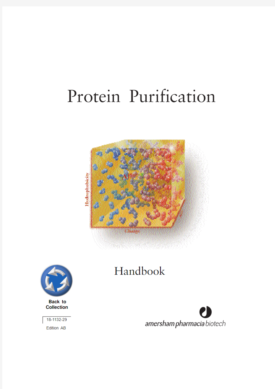

Protein Purification
Handbook 18-1132-29
Edition AB
Back to
Collection
HiTrap, Sepharose, STREAMLINE, Sephadex, MonoBeads, Mono Q,
Mono S, MiniBeads, RESOURCE, SOURCE, Superdex, Superose, HisTrap, HiLoad, HiPrep, INdEX, BPG, BioProcess, FineLINE, MabTrap, MAbAssistant, Multiphor, FPLC, PhastSystem and ?KTA are trademarks of Amersham Pharmacia Biotech Limited
or its subsidiaries.
Amersham is a trademark of Nycomed Amersham plc
Pharmacia and Drop Design are trademarks of Pharmacia & Upjohn Inc Coomassie is a trademark of ICI plc
All goods and services are sold subject to the terms and conditions of sale of the company within the Amersham Pharmacia Biotech group which supplies them. A copy of these terms and conditions of sale is available on request.
? Amersham Pharmacia Biotech AB 1999-All rights reserved.
Amersham Pharmacia Biotech AB
SE-751 84 Uppsala Sweden
Amersham Pharmacia Biotech UK Limited Amersham Place Little Chalfont Buckinghamshire England HP7 9NA Amersham Pharmacia Biotech Inc
800 Centennial Avenue PO Box 1327 Piscataway NJ 08855 USA
Protein Purification Handbook
Contents Introduction (7)
Chapter 1
Purification Strategies - A Simple Approach (9)
Preparation (10)
Three Phase Purification Strategy (10)
General Guidelines for Protein Purification (12)
Chapter 2 Preparation (13)
Before You Start (13)
Sample Extraction and Clarification (16)
Chapter 3
Three Phase Purification Strategy (19)
Principles (19)
Selection and Combination of Purification Techniques (20)
Sample Conditioning (26)
Chapter 4
Capture (29)
Chapter 5
Intermediate Purification (37)
Chapter 6
Polishing (40)
Chapter 7
Examples of Protein Purification Strategies (45)
Three step purification of a recombinant enzyme (45)
Three step purification of a recombinant antigen binding fragment (49)
Two step purification of a monoclonal antibody (54)
One step purification of an integral membrane protein (57)
Chapter 8
Storage Conditions (61)
Extraction and Clarification Procedures (62)
Chapter 9
Principles and Standard Conditions for Purification Techniques (73)
Ion exchange (IEX) (73)
Hydrophobic interaction (HIC) (79)
Affinity (AC) (85)
Gel filtration (GF) (88)
Reversed phase (RPC) (92)
Expanded bed adsorption (EBA) (95)
Introduction
The development of techniques and methods for protein purification has been an essential pre-requisite for many of the advancements made in biotechnology. This booklet provides advice and examples for a smooth path to protein purification. Protein purification varies from simple one-step precipitation procedures to large scale validated production processes. Often more than one purification step is necessary to reach the desired purity. The key to successful and efficient protein purification is to select the most appropriate techniques, optimise their performance to suit the requirements and combine them in a logical way to maximise yield and minimise the number of steps required.
Most purification schemes involve some form of chromatography. As a result chromatography has become an essential tool in every laboratory where protein purification is needed. The availability of different chromatography techniques with different selectivities provides a powerful combination for the purification of any biomolecule.
Recombinant DNA developments over the past decade have revolutionised the production of proteins in large quantities. Proteins can even be produced in forms which facilitate their subsequent chromatographic purification. However, this has not removed all challenges. Host contaminants are still present and problems related to solubility, structural integrity and biological activity can still exist. Although there may appear to be a great number of parameters to consider, with a few simple guidelines and application of the Three Phase Purification Strategy the process can be planned and performed simply and easily, with only a basic knowledge of the details of chromatography techniques.
7
8
Chapter 1
Purification Strategies
- a simple approach
Apply a systematic approach to development of a purification strategy. The first step is to describe the basic scenario for the purification. General considerations answer questions such as: What is the intended use of the product? What kind of starting material is available and how should it be handled? What are the purity issues in relation to the source material and intended use of the final product? What has to be removed? What must be removed completely? What will be the final scale of purification? If there is a need for scale-up, what consequences will this have on the chosen purification techniques? What are the economical constraints and what resources and equipment are available?
Most purification protocols require more than one step to achieve the desired level of product purity. This includes any conditioning steps necessary to transfer the product from one technique into conditions suitable to perform the next technique. Each step in the process will cause some loss of product. For example, if a yield of 80% in each step is assumed, this will be reduced to only 20% overall yield after 8 processing steps as shown in Figure 1. Consequently, to reach the targets for yield and purity with the minimum number of steps and the simplest possible design, it is not efficient to add one step to another until purity requirements have been fulfilled. Occasionally when a sample is readily available purity can be achieved by simply adding or repeating steps. However, experience shows that, even for the most challenging applications, high purity and yield can be achieved efficiently in fewer than four well-chosen and optimised purification steps. Techniques should be organised in a logical sequence to avoid the need for conditioning steps and the chromatographic techniques selected appropriately to use as few purification steps as possible.
Limit the number of steps in a purification procedure
9
10
Fig.1.Yields from multi-step purifications.
Preparation
The need to obtain a protein, efficiently, economically and in sufficient purity and quantity, applies to every purification. It is important to set objectives for purity,quantity and maintenance of biological activity and to define the economical and time framework for the work. All information concerning properties of the target protein and contaminants will help during purification development. Some simple experiments to characterise the sample and target molecule are an excellent investment. Development of fast and reliable analytical assays is essential to follow the progress of the purification and assess its effectiveness. Sample
preparation and extraction procedures should be developed prior to the first chromatographic purification step.
With background information, assays and sample preparation procedures in place the Three Phase Purification Strategy can be considered.
Three Phase Purification Strategy Imagine the purification has three phases Capture, Intermediate
Purification and Polishing.
In the Three Phase Strategy specific objectives are assigned to each step within the process:
In the capture phase the objectives are to isolate, concentrate and stabilise the target product.
During the intermediate purification phase the objective is to remove most of the bulk impurities such as other proteins and nucleic acids, endotoxins and viruses.In the polishing phase the objective is to achieve high purity by removing any remaining trace impurities or closely related substances.
The selection and optimum combination of purification techniques for Capture,Intermediate Purification and Polishing is crucial to ensure fast method development, a shorter time to pure product and good economy.
10
80
60
40
20
12345678Number of steps 95% / step
90% / step 85% / step 80% / step 75% / step
Yield (%)
The final purification process should ideally consist of sample preparation, including extraction and clarification when required, followed by three major purification steps, as shown in Figure 2. The number of steps used will always depend upon the purity requirements and intended use for the protein.
Fig. 2.Preparation and the Three Phase Purification Strategy
11
Guidelines for Protein Purification
The guidelines for protein purification shown here can be applied to any purification process and are a suggestion as to how a systematic approach can be applied to the development of an effective purification strategy. As a reminder these guidelines will be highlighted where appropriate throughout the following chapters.
Define objectives
for purity, activity and quantity required of final product to avoid over or under developing a method
Define properties of target protein and critical impurities
to simplify technique selection and optimisation
Develop analytical assays
for fast detection of protein activity/recovery and to work efficiently
Minimise sample handling at every stage
to avoid lengthy procedures which risk losing activity/reducing recovery Minimise use of additives
additives may need to be removed in an extra purification step or may interfere with activity assays
Remove damaging contaminants early
for example, proteases
Use a different technique at each step
to take advantage of sample characteristics which can be used for separation (size, charge, hydrophobicity, ligand specificity)
Minimise number of steps
extra steps reduce yield and increase time, combine steps logically
KEEP IT SIMPLE!
12
Chapter 2
Preparation
Before You Start
The need to obtain a protein, efficiently, economically and in sufficient purity and quantity, applies to any purification, from preparation of an enriched protein extract for biochemical characterisation to large scale production of a therapeutic recombinant protein. It is important to set objectives for purity and quantity, maintenance of biological activity and economy in terms of money and time. Purity requirements must take into consideration the nature of the source material, the intended use of the final product and any special safety issues. For example, it is important to differentiate between contaminants which must be removed and those which can be tolerated. Other factors can also influence the prioritisation of objectives. High yields are usually a key objective, but may be less crucial in cases where a sample is readily available or product is required only in small quantities. Extensive method development may be impossible without resources such as an ?KTA?design chromatography system. Similarly, time pressure combined with a slow assay turnaround will steer towards less extensive scouting and optimisation. All information concerning properties of the target protein and contaminants will help during purification development, allowing faster and easier technique selection and optimisation, and avoiding conditions which may inactivate the target protein.
Development of fast and reliable analytical assays is essential to follow the progress of the purification and assess effectiveness (yield, biological activity, recovery).
Define objectives
Goal:To set minimum objectives for purity and quantity, maintenance of biological activity and economy in terms of money and time.
Define purity requirements according to the final use of the product. Purity requirement examples are shown below.
Extremely high > 99%Therapeutic use, in vivo studies
High 95- 99 %X-ray crystallography and most physico-chemical
characterisation methods
Moderate < 95 %Antigen for antibody production
N-terminal sequencing
13
Identify 'key' contaminants
Identify the nature of possible remaining contaminants as soon as
possible.
The statement that a protein is >95% pure (i.e. target protein constitutes 95% of total protein) is far from a guarantee that the purity is sufficient for an intended application. The same is true for the common statement "the protein was homogenous by Coomassie? stained SDS-PAGE". Purity of 95% may be acceptable if the remaining 5% consists of harmless impurities. However, even minor impurities which may be biologically active could cause significant problems in both research and therapeutic applications. It is therefore important to differentiate between contaminants which must be removed completely and those which can be reduced to acceptable levels. Since different types of starting material will contain different contaminant profiles they will present different contamination problems.
It is better to over-purify than to under-purify.
Although the number of purification steps should be minimised, the
quality of the end product should not be compromised. Subsequent results might be questioned if sample purity is low and contaminants are unknown.
Contaminants which degrade or inactivate the protein or interfere with
analyses should be removed as early as possible.
The need to maintain biological activity must be considered at every stage during purification development. It is especially beneficial if proteases are removed and target protein transferred into a friendly environment during the first step.
Economy is a very complex issue. In commercial production the time to market can override issues such as optimisation for recovery, capacity or speed. Robustness and reliability are also of great concern since a batch failure can have major consequences.
It may be necessary to use analytical techniques targetted towards specific conta-minants in order to demonstrate that they have been removed to acceptable levels. 14
Define properties of target protein and critical impurities Goal:To determine a 'stability window' for the target protein for easier selection and optimisation of techniques and to avoid protein inactivation during purification.
Check target protein stability window for at least pH and ionic strength. All information concerning the target protein and contaminant properties will help to guide the choice of separation techniques and experimental conditions for purification. Database information for the target, or related proteins, may give size, isoelectric point (pI) and hydrophobicity or solubility data. Native one and two dimensional PAGE can indicate sample complexity and the properties of the target protein and major contaminants. Particularly important is a knowledge of the stability window of the protein so that irreversible inactivation is avoided. It
is advisable to check the target protein stability window for at least pH and ionic strength. Table 1 shows how different target protein properties can affect a purification strategy.
Table 1.Protein properties and their effect on development of purification strategies. Sample and target protein properties Influence on purification strategy
Temperature stability Need to work rapidly at lowered temperature
pH stability Selection of buffers for extraction and purification
Selection of conditions for ion exchange, affinity or
reversed phase chromatography
Organic solvents stability Selection of conditions for reversed phase
chromatography
Detergent requirement Consider effects on chromatographic steps and the need
for detergent removal. Consider choice of detergent.
Salt (ionic strength)Selection of conditions for precipitation techniques and
hydrophobic interaction chromatography
Co-factors for stability or activity Selection of additives, pH, salts, buffers
Protease sensitivity Need for fast removal of proteases or addition of
inhibitors
Sensitivity to metal ions Need to add EDTA or EGTA in buffers
Redox sensitivity Need to add reducing agents
Molecular weight Selection of gel filtration media
Charge properties Selection of ion exchange conditions
Biospecific affinity Selection of ligand for affinity medium
Post translational modifications Selection of group specific affinity medium Hydrophobicity Selection of medium for hydrophobic interaction
chromatography
15
Develop analytical assays
Goal:To follow the progress of a purification, to assess effectiveness (yield, biological activity, recovery) and to help during optimisation.
Select assays which are fast and reliable.
To progress efficiently during method development the effectiveness of each step should be assessed. The laboratory should have access to the following assays:? A rapid, reliable assay for the target protein
? Purity determination
? Total protein determination
? Assays for impurities which must be removed
The importance of a reliable assay for the target protein cannot be over- emphasised. When testing chromatographic fractions ensure that the buffers used for separation do not interfere with the assay. Purity of the target protein is most often estimated by SDS-PAGE, capillary electrophoresis, reversed phase chromatography or mass spectrometry. Lowry or Bradford assays are used most frequently to determine the total protein.
The Bradford assay is particularly suited to samples where there is a high lipid content which may interfere with the Lowry assay.
For large scale protein purification the need to assay for target proteins and critical impurities is often essential. In practice, when a protein is purified for research purposes, it is too time consuming to identify and set up specific assays for harmful contaminants. A practical approach is to purify the protein to a certain level, and then perform SDS-PAGE after a storage period to check for protease cleavage. Suitable control experiments, included within assays for
bio-activity, will help to indicate if impurities are interfering with results.
Sample Extraction and Clarification Minimise sample handling
Minimise use of additives
Remove damaging contaminants early
Definition:Primary isolation of target protein from source material.
Goal:Preparation of a clarified sample for further purification. Removal of particulate matter or other contaminants which are not compatible with chromatography.
16
The need for sample preparation prior to the first chromatographic step is dependent upon sample type. In some situations samples may be taken directly to the first capture step. For example cell culture supernatant can be applied directly to a suitable chromatographic matrix such as Sepharose? Fast Flow and may require only a minor adjustment of the pH or ionic strength. However, it is most often essential to perform some form of sample extraction and clarification procedure.
If sample extraction is required the chosen technique must be robust and suitable for all scales of purification likely to be used. It should be noted that a technique such as ammonium sulphate precipitation, commonly used in small scale, may be unsuitable for very large scale preparation. Choice of buffers and additives must be carefully considered if a purification is to be scaled up. In these cases inexpensive buffers, such as acetate or citrate, are preferable to the more complex compositions used in the laboratory. It should also be noted that dialysis and other common methods used for adjustment of sample conditions are unsuitable for very large or very small samples.
For repeated purification, use an extraction and clarification technique
that is robust and able to handle sample variability. This ensures a
reproducible product for the next purification step despite variability in
starting material.
Use additives only if essential for stabilisation of product or improved
extraction. Select those which are easily removed. Additives may need to
be removed in an extra purification step.
Use pre-packed columns of Sephadex? G-25 gel filtration media, for
rapid sample clean-up at laboratory scale, as shown in Table 2.
Table 2.Pre-packed columns for sample clean-up.
Pre-packed column Sample volume Sample volume Code No.
loading per run recovery per run
HiPrep?Desalting 26/10 2.5 -15 ml7.5 - 20 ml17-5087-01
HiTrap Desalting0.25 - 1.5 ml 1.0 - 2.0 ml17-1408-01
Fast Desalting PC 3.2/100.05 - 0.2 ml0.2 - 0.3 ml17-0774-01
PD-10 Desalting 1.5 - 2.5 ml 2.5 - 3.5 ml17-0851-01 Sephadex G-25 gel filtration media are used at laboratory and production scale for sample preparation and clarification of proteins >5000. Sample volumes of up to 30%, or in some cases, 40% of the total column volume are loaded. In a single step, the sample is desalted, exchanged into a new buffer, and low molecular weight materials are removed. The high volume capacity, relative insensitivity to sample concentration, and speed of this step enable very large sample volumes to be processed rapidly and efficiently. Using a high sample volume load results in a separation with minimal sample dilution (approximately 1:1.4). Chapter 8 contains further details on sample storage, extraction and clarification procedures.
17
Sephadex G-25 is also used for sample conditioning i.e. rapid adjustment of pH, buffer exchange and desalting between purification steps.
Sephadex G-25 gel filtration
For fast group separations between high and low molecular weight substances Typical flow velocity 60 cm/h (Sephadex G-25 SuperFine, Sephadex G-25 Fine), 150 cm/h (Sephadex G-25 Medium).
If large sample volumes will be handled or the method scaled-up in the future, consider using STREAMLINE? expanded bed adsorption. This technique is particularly suited for large scale recombinant protein and monoclonal antibody purification. The crude sample containing particles can be applied to the expanded bed without filtration or centrifugation. STREAMLINE adsorbents are specially designed for use in STREAMLINE columns. Together they enable the high flow rates needed for high productivity in industrial applications of fluidised beds. The technique requires no sample clean up and so combines sample preparation and capture in a single step. Crude sample is applied to an expanded bed STREAMLINE media. Target proteins are captured whilst cell debris, cells, particulate matter, whole cells, and contaminants pass through. Flow is reversed and the target proteins are desorbed in the elution buffer.
STREAMLINE (IEX, AC, HIC)
For sample clean-up and capture direct from crude sample.
STREAMLINE adsorbents are designed to handle feed directly from both fermentation homogenate and crude feedstock from cell culture/fermentation at flow velocities of 200 - 500 cm/h, according to type and application.
Particle size: 200 μm
Note:cm/h: flow velocity (linear flow rate) = volumetric flow rate/cross sectional area of column.
18
Chapter 3
Three Phase Purification Strategy
Principles
With background information, assays, and sample preparation and extraction procedures in place the Three Phase Purification Strategy can be applied (Figure 3). This strategy is used as an aid to the development of purification processes for therapeutic proteins in the pharmaceutical industry and is equally efficient as an aid when developing purification schemes in the research laboratory.
Fig. 3.Preparation and the Three Phase Purification Strategy.
Assign a specific objective to each step within the purification process.
In the Three Phase Strategy a specific objective is assigned to each step. The purification problem associated with a particular step will depend greatly upon the properties of the starting material. Thus, the objective of a purification step will vary according to its position in the process i.e. at the beginning for isolation of product from crude sample, in the middle for further purification of partially purified sample, or at the end for final clean up of an almost pure product.
The Three Phase Strategy ensures faster method development, a shorter time to pure product and good economy.
In the capture phase the objectives are to isolate, concentrate and stabilise the target product. The product should be concentrated and transferred to an environment which will conserve potency/activity. At best, significant removal of other critical contaminants can also be achieved.
19
During the intermediate purification phase the objectives are to remove most of the bulk impurities,such as other proteins and nucleic acids, endotoxins and viruses.
In the polishing phase most impurities have already been removed except for trace amounts or closely related substances. The objective is to achieve final purity.
It should be noted that this Three Phase Strategy does not mean that all strategies must have three purification steps. For example, capture and intermediate purification may be achievable in a single step, as may intermediate purification and polishing. Similarly, purity demands may be so low that a rapid capture step is sufficient to achieve the desired result, or the purity of the starting material may be so high that only a polishing step is needed. For purification of therapeutic proteins a fourth or fifth purification step may be required to fulfil the highest purity and safety demands.
The optimum selection and combination of purification techniques for Capture, Intermediate Purification and Polishing is crucial for an efficient purification process.
Selection and Combination of
Purification Techniques
Minimise sample handling
Minimise number of steps
Use different techniques at each step
Goal:Fastest route to a product of required purity.
For any chromatographic separation each different technique will offer different performance with respect to recovery, resolution, speed and capacity. A technique can be optimised to focus on one of these parameters, for example resolution, or to achieve the best balance between two parameters, such as speed and capacity.
A separation optimised for one of these parameters will produce results quite different in appearance from those produced using the same technique, but focussed on an alternative parameter. See, for example, the results shown on page 49 where ion exchange is used for a capture and for a polishing step.
20
Select a technique to meet the objectives for the purification step. Capacity,in the simple model shown, refers to the amount of target protein loaded during purification. In some cases the amount of sample which can be loaded may be limited by volume (as in gel filtration) or by large amounts of contaminants rather than the amount of the target protein.
Speed is of the highest importance at the beginning of a purification where contaminants such as proteases must be removed as quickly as possible. Recovery becomes increasingly important as the purification proceeds because of the increased value of the purified product. Recovery is influenced by destructive processes in the sample and unfavourable conditions on the column. Resolution is achieved by the selectivity of the technique and the efficiency of the chromatographic matrix to produce narrow peaks. In general, resolution is most difficult to achieve in the final stages of purification when impurities and target protein are likely to have very similar properties.
Every technique offers a balance between resolution, speed, capacity and recovery and should be selected to meet the objectives for each purification step. In general, optimisation of any one of these four parameters can only be achieved at the expense of the others and a purification step will be a compromise. The importance of each parameter will vary depending on whether a purification step is used for capture, intermediate purification or polishing. This will steer the optimisation of the critical parameters, as well as the selection of the most suitable media for the step.
Proteins are purified using chromatographic purification techniques which separate according to differences in specific properties, as shown in Table 3. Table 3.Protein properties used during purification.
Protein property Technique
Charge Ion exchange (IEX)
Size Gel filtration (GF)
Hydrophobicity Hydrophobic interaction (HIC),
reversed phase (RPC)
Biorecognition (ligand specificity)Affinity (AC)
Charge, ligand specificity or hydrophobicity Expanded bed adsorption (EBA) follows the
principles of AC, IEX or HIC
21
Choose logical combinations of purification techniques based on the main benefits of the technique and the condition of the sample at the beginning
or end of each step.
Minimise sample handling between purification steps by combining
techniques to avoid the need for sample conditioning.
A guide to the suitability of each purification technique for the stages in the Three Phase Purification Strategy is shown in Table 4.
Table 4.Suitability of purification techniques for the Three Phase Purification Strategy Avoid additional sample conditioning steps.
The product should be eluted from the first column in conditions suitable for the start conditions of the next column.
The start conditions and end conditions for the techniques are shown in Table 4. For example, if the sample has a low ionic strength it can be applied to an IEX column. After elution from IEX the sample will usually be in a high ionic strength buffer and can be applied to a HIC column (if necessary the pH can be adjusted and further salt can be added). In contrast, if sample is eluted from a HIC column, it is likely to be in high salt and will require dilution or a buffer exchange step in order to further decrease the ionic strength to a level suitable for IEX. Thus it is more straightforward to go from IEX to HIC than vice-versa.
step. The salt concentration and the total sample volume will be significantly reduced after elution from the HIC column. Dilution of the fractionated sample or rapid buffer exchange using a Sephadex G-25 desalting column will prepare it for the next IEX or AC step.
22
蛋白质纯化的方法 蛋白质的分离纯化方法很多,主要有: (一)根据蛋白质溶解度不同的分离方法 1、蛋白质的盐析 中性盐对蛋白质的溶解度有显著影响,一般在低盐浓度下随着盐浓度升高,蛋白质的溶解度增加,此称盐溶;当盐浓度继续升高时,蛋白质的溶解度不同程度下降并先后析出,这种现象称盐析,将大量盐加到蛋白质溶液中,高浓度的盐离子(如硫酸铵的SO4和NH4)有很强的水化力,可夺取蛋白质分子的水化层,使之“失水”,于是蛋白质胶粒凝结并沉淀析出。盐析时若溶液pH在蛋白质等电点则效果更好。由于各种蛋白质分子颗粒大小、亲水程度不同,故盐析所需的盐浓度也不一样,因此调节混合蛋白质溶液中的中性盐浓度可使各种蛋白质分段沉淀。 影响盐析的因素有:(1)温度:除对温度敏感的蛋白质在低温(4度)操作外,一般可在室温中进行。一般温度低蛋白质溶介度降低。但有的蛋白质(如血红蛋白、肌红蛋白、清蛋白)在较高的温度(25度)比0度时溶解度低,更容易盐析。(2)pH值:大多数蛋白质在等电点时在浓盐溶液中的溶介度最低。(3)蛋白质浓度:蛋白质浓度高时,欲分离的蛋白质常常夹杂着其他蛋白质地一起沉淀出来(共沉现象)。因此在盐析前血清要加等量生理盐水稀释,使蛋白质含量在2.5-3.0%。 蛋白质盐析常用的中性盐,主要有硫酸铵、硫酸镁、硫酸钠、氯化钠、磷酸钠等。其中应用最多的硫酸铵,它的优点是温度系数小而溶解度大(25度时饱和溶液为4.1M,即767克/升;0度时饱和溶解度为3.9M,即676克/升),在这一溶解度范围内,许多蛋白质和酶都可以盐析出来;另外硫酸铵分段盐析效果也比其他盐好,不易引起蛋白质变性。硫酸铵溶液的pH常在4.5-5.5之间,当用其他pH值进行盐析时,需用硫酸或氨水调节。 蛋白质在用盐析沉淀分离后,需要将蛋白质中的盐除去,常用的办法是透析,即把蛋白质溶液装入秀析袋内(常用的是玻璃纸),用缓冲液进行透析,并不断的更换缓冲液,因透析所需时间较长,所以最好在低温中进行。此外也可用葡萄糖凝胶G-25或G-50过柱的办法除盐,所用的时间就比较短。
AKTA蛋白纯化系统操作 AKTA蛋白纯化系统是当前蛋白纯化工作经常用到的一组设备,自动化程度很高。AKTA系统依据不同的配置,可以分为AKTA EXPLORER、AKTA PILOT、AKTA PURIFIER等多种型号的设备。以下以AKTA EXPLORER为例简单介绍AKTA蛋白纯化系统的一般操作。 1、认识AKTA。 AKTA explorer 是为方法开拓及研究应用而设计的全自动液相色谱系统。该色谱系统的分离装置有三个主要组件,在底部平台的左侧整齐堆起(Fig 1)。它们是: FIG 1、AKTA EXPLORER主机 ? Pump-900 为双通道高效梯度泵系列。在AKTAexplorer 100,流速范围0.01-100 ml/min,压力高达10 Mpa(泵名为P-901)。在AKTA explore10,流速范围0.001-10 ml/min,压力高达25 Mpa(泵名为P-903)。 ? Monitor UV-900,同时监控190-700 nm 范围内高达3 个波长的多波长紫外-可见(UV-Vis)监测器。(针对部分AKTA PURIFIER机型,尚有UPC-900监测器可供选择,光源为汞灯光源,一次可以监控一个波长,安装滤光片后,可以在选择的波长范围内进行切换。)? Monitor pH/C-900,在线电导和pH 监测的组合监测器。 Fig 2、AKTA EXPLORER硬件模式图
AKTA EXPLORER系统的主要组成部件可以用模式图表示(Fig 2)。组成部件,如混合器、柱及不同的阀安装在右边部分。打开装阀的门可全部看到。柱被挂在装阀的门的外侧。 分离装置由UNICORN 软件控制。软件安装于一独立的电脑主机之中,在电脑与色谱系统之间的通信由数据采集装置CU950进行控制。 2、一般操作 2.1 开机 按位于底部平台前左侧的ON/OFF 按钮,打开色谱系统,然后打开电脑电源。待仪器自检完毕(CU950上面的3个指示灯完全点亮并不闪烁)。双击桌面上UNICORN图标,进入操作界面。UNICORN的操作界面分为四个窗口(Fig 3) Fig 3、Unicorn的操作界面 2.2准备工作溶液和样品 所有的工作溶液和样品必须经过0.45μm的滤膜过滤,样品也可高速离心后取上清备用。当缓冲液中含有有机溶剂(如乙腈、甲醇),需在使用前用低频超声脱气10min。 2.3清洗及管道准备 首先将A泵的进液管道(A1)放入缓冲液或平衡液中,将B泵的进液管道(B1)放入高盐溶液中,在system control窗口点击工具栏内的manual,选择pump→pump wash explorer,选中A1,B1管道为ON,execute。泵清洗将自动结束。(Fig 4) Fig 4、AKTA Explorer的泵清洗操作 2.4安装层析柱
蛋白质分离纯化的一般程序可分为以下几个步骤: (一)材料的预处理及细胞破碎 分离提纯某一种蛋白质时,首先要把蛋白质从组织或细胞中释放出来并保持原来的天然状态,不丧失活性。所以要采用适当的方法将组织和细胞破碎。常用的破碎组织细胞的方法有: 1. 机械破碎法 这种方法是利用机械力的剪切作用,使细胞破碎。常用设备有,高速组织捣碎机、匀浆器、研钵等。 2. 渗透破碎法 这种方法是在低渗条件使细胞溶胀而破碎。 3. 反复冻融法 生物组织经冻结后,细胞内液结冰膨胀而使细胞胀破。这种方法简单方便,但要注意那些对温度变化敏感的蛋白质不宜采用此法。 4. 超声波法 使用超声波震荡器使细胞膜上所受张力不均而使细胞破碎。 5. 酶法 如用溶菌酶破坏微生物细胞等。 (二)蛋白质的抽提 通常选择适当的缓冲液溶剂把蛋白质提取出来。抽提所用缓冲液的pH、离子强度、组成成分等条件的选择应根据欲制备的蛋白质的性质而定。如膜蛋白的抽提,抽提缓冲液中一般要加入表面活性剂(十二烷基磺酸钠、tritonX-100 等),使膜结构破坏,利于蛋白质与膜分离。在抽提过程中,应注意温度,避免剧烈搅拌等,以防止蛋白质的变性。(三)蛋白质粗制品的获得选用适当的方法将所要的蛋白质与其它杂蛋白分离开来。比较方便的有效方法是根据蛋白质溶解度的差异进行的分离。常用的有下列几种方法: 1.等电点沉淀法不同蛋白质的等电点不同,可用等电点沉淀法使它们相互分离。 2.盐析法 不同蛋白质盐析所需要的盐饱和度不同,所以可通过调节盐浓度将目的蛋白沉淀析出。被盐析沉淀下来的蛋白质仍保持其天然性质,并能再度溶解而不变性。 3.有机溶剂沉淀法 中性有机溶剂如乙醇、丙酮,它们的介电常数比水低。能使大多数球状蛋白质在水溶液中的溶解度降低,进而从溶液中沉淀出来,因此可用来沉淀蛋白质。此外,有机溶剂会破坏蛋白质表面的水化层,促使蛋白质分子变得不稳定而析出。由于有机溶剂会使蛋白质变性,使用该法时,要注意在低温下操作,选择合适的有机溶剂浓度。 (四)样品的进一步分离纯化
蛋白质的提取和纯化-- 选择分离材料及预处理蛋白质的提取和纯化-- 选择分离材料及预处理 以蛋白质和结构与功能为基础,从分子水平上认识生命现象,已经成为现代生物学发展的主要方向,研究蛋白质,首先要得到高度纯化并具有生物活性的目的物质。 蛋白质的制备工作涉及物理、化学和生物等各方面知识,但基本原理不外乎两方面。一是得用混合物中几个组分分配率的差别,把它们分配到可用机械方法分离的两个或几个物相中,如盐析,有机溶剂提取,层析和结晶等;二是将混合物置于单一物相中,通过物理力场的作用使各组分分配于来同区域而达到分离目的,如电泳,超速离心,超滤等。在所有这些方法的应用中必须注意保存生物大分子的完整性,防止酸、硷、高温,剧烈机械作用而导致所提物质生物活性的丧失。 蛋白质的制备一般分为以下四个阶段:选择材料和预处理,细胞的破碎及细胞器的分离,提取和纯化,浓细、干燥和保存。 微生物、植物和动物都可做为制备蛋白质的原材料,所选用的材料主要依据实验目的来确定。对于微生物,应注意它的生长期,在微生物的对数生长期,酶和核酸的含量较高,可以获得高产量,以微生物为材料时有两种情况:( 1 )得用微生物菌体分泌到培养基中的代谢产物和胞外酶等;(2)利用菌体 含有的生化物质,如蛋白质、核酸和胞内酶等。植物材料必须经过去壳,脱脂并注意植物品种和生长发育状况不同,其中所含生物大分子的量变化很大,另外与季节性关系密切。对动物组织,必须选择有效成份含量丰富的脏器组织为原材料,先进行绞碎、脱脂等处理。另外,对预处理好的材料,若不立即进行实验,应冷冻保存,对于易分解的生物大分子应选用新鲜材料制备。 细胞的破碎 1、高速组织捣碎:将材料配成稀糊状液,放置于筒内约1/3 体积,盖紧筒盖,将调速器先拨至最慢处, 开动开关后,逐步加速至所需速度。此法适用于动物内脏组织、植物肉质种子等。 2、玻璃匀浆器匀浆:先将剪碎的组织置于管中,再套入研杆来回研磨,上下移动,即可将细胞研碎,此法细胞破碎程度比高速组织捣碎机为高,适用于量少和动物脏器组织。 3、超声波处理法:用一定功率的超声波处理细胞悬液,使细胞急剧震荡破裂,此法多适用于微生物材料, 用大肠杆菌制备各种酶,常选用50-100 毫克菌体/毫升浓度,在1KG 至10KG 频率下处理10-15 分钟,此法的缺点是在处理过程会产生大量的热,应采取相应降温措施。对超声波敏感和核酸应慎用。 4、反复冻融法:将细胞在-20 度以下冰冻,室温融解,反复几次,由于细胞内冰粒形成和剩余细胞液的盐浓度增高引起溶胀,使细胞结构破碎。 5、化学处理法:有些动物细胞,例如肿瘤细胞可采用十二烷基磺酸钠(SDS)、去氧胆酸钠等细胞膜破 坏,细菌细胞壁较厚,可采用溶菌酶处理效果更好。 无论用哪一种方法破碎组织细胞,都会使细胞内蛋白质或核酸水解酶释放到溶液中,使大分子生物 降解,导致天然物质量的减少,加入二异丙基氟磷酸(DFP)可以抑制或减慢自溶作用;加入碘乙酸可以
随着分子生物学的发展,越来越多的科研人员熟练掌握了分子生物学的各种试验技术,并研制成套试剂盒,使基因克隆表达变得越来越容易lIl。但分子生物学的上游工作往往并非是最终目的,分子克隆与表达的关键是要拿到纯的表达产物,以研究其生物学作用,或者大量生产出可用于疾病治疗的生物制品。相对与上游工作来说,分子克隆的下游工作显得更难,蛋白纯化工作非常复杂,除了要保证纯度外,蛋白产品还必须保持其生物学活性。纯化工艺必须能够每次都能产生相同数量和质量的蛋白,重复性良好。这就要求应用适应性非常强的方法而不是用能得到纯蛋白的最好方法去纯化蛋白。在实验室条件下的好方法却可能在大规模生产应用中失败,因为后者要求规模化,且在每日的应用中要有很好的重复性。本文综述了蛋白质纯化的基本原则和各种蛋白纯化技术的原理、优点及局限性,以期对蛋白纯化的方法选择及整体方案的制定提供一定的指导。 1 蛋白纯化的一般原则 蛋白纯化要利用不同蛋白间内在的相似性与差异,利用各种蛋白间的相似性来除去非蛋白物质的污染,而利用各蛋白质的差异将目的蛋白从其他蛋白中纯化出来。每种蛋白间的大小、形状、电荷、疏水性、溶解度和生物学活性都会有差异,利用这些差异可将蛋白从混合物如大肠杆菌裂解物中提取出来得到重组蛋白。蛋白的纯化大致分为粗分离阶段和精细纯化阶段二个阶段。粗分离阶段主要将目的蛋白和其他细胞成分如DNA、RNA等分开,由于此时样本体积大、成分杂,要求所用的树脂高容量、高流速,颗粒大、粒径分布宽.并可以迅速将蛋白与污染物分开,防止目的蛋白被降解。精细纯化阶段则需要更高的分辨率,此阶段是要把目的蛋白与那些大小及理化性质接近的蛋白区分开来,要用更小的树脂颗粒以提高分辨常用的离子交换柱和疏水柱,应用时要综合考虑树脂的选择性和柱效两个因素。选择性指树脂与目的蛋白结合的特异性,柱效则是指蛋白的各成分逐个从树脂上集中洗脱的能力,洗脱峰越窄,柱效越好。仅有好的选择性,洗脱峰太宽,蛋白照样不能有效分离。 2.各种蛋白纯化方法及优缺点 2.1蛋白沉淀蛋白能溶于水是因为其表面有亲水性氨基酸。在蛋白质的等电点处若溶液的离子强度特别高或特别低,蛋白则倾向于从溶液中析出。硫酸铵是沉淀蛋白质最常用的盐,因为它在冷的缓冲液中溶解性好,冷的缓冲液有利于保护蛋白的活性。硫酸
蛋白纯化系统操作流程 一、蛋白的诱导:蛋白原核表达 1、取菌种接种于含Amp LB固体培养基中(分区划线),37℃培养过夜; 2、挑取单克隆接种于5ml含Amp LB液体培养基中,37℃振摇过夜; 3、从过夜培养物中取2ml接种于100ml Amp LB液体培养基中,振摇2h(留样1ml); 4、加入一定终浓度IPTG,37℃诱导表达4h(留样1ml),离心,弃上清收集细菌。 存入4℃。 二、蛋白表达状态分析(可溶性or包涵体表达) 取少量(1ml)诱导菌体沉淀,加入不含变性剂(如盐酸胍,尿素等)PBS(150μl),超声裂解。分离上清和沉淀,分别SDS-PAGE电泳。 三、蛋白的纯化 纯化前准备 1.推荐在中性至弱碱性条件下(pH 7-8)结合重组蛋白。磷酸盐buffer是常用的缓冲液, Tric-Cl在一般情况下可用,但要注意它会降低结合强度。 2.避免在buffer中包含EDTA或柠檬酸盐等螯合剂 3.若重组蛋白以包涵体形式表达,在所有的buffer中添加6 M 盐酸胍或8 M 尿素 注: 1.包含尿素的样品可直接进行SDS-PAGE分析,若样品中包含盐酸胍,在SDS-PAGE前则 需先用含有尿素的buffer进行透析 2.除利用咪唑洗脱蛋白,其它方法,如低pH 值法等可被应用,详见说明书 Bingding buffer 中咪唑的浓度 在洗涤时所用的Bingding buffer 中咪唑浓度越大,重组蛋白纯度越高。但过高的咪唑浓度将导致蛋白的洗脱。合适的咪唑浓度需要优化。 Buffer 的准备
所用的水及化学物质须是高纯度的。Buffer 在使用前需经0.45 μm滤膜过滤 所用高纯度的咪唑需在280nm 处无吸光度或吸光度极低 推荐buffer Bingding buffer:20 mM 磷酸盐 0.5 M NaCl 20- 40 mM 咪唑pH 7.4 (咪唑浓度是蛋白依赖的,可变!)Elution buffer :20 mM 磷酸盐 0.5 M NaCl 500 mM 咪唑pH 7.4 (咪唑浓度是蛋白依赖的,可变!) 样品准备 样品需被充分溶解。过柱前经0.45 μm滤膜过滤。样品以pH 7.4 binding buffer 溶解。勿用强酸强碱调节pH 值,否则将可能导致沉淀。 重力纯化法Ni-NTA Column 准备 1. 温和地颠倒瓶中的Ni-NTA Agarose 数次。 2. 吸取2ml的树脂加入15ml离心管中,使树脂在重力(5–10 minutes)或低速离心(5 minute at 500 × g),轻柔的吸出上清。 3. 加入5ml的无菌蒸馏水,温和的颠倒柱子3min,离心5 minute at 500 × g,轻柔的吸出上清。 4. 用bingding buffer 重复第3步。 5. 在Ni 柱中加入等体积的bingding buffer,制成50%的slurry 样品与Ni 柱结合 1.每1ml 50%的slurry中加入4ml 的样品。1ml 50%的slurry 可结合20mg His-蛋白 2.将混合物室温,低速振荡孵育1h Buffer 洗涤及洗脱 1.离心5 minute at 500 × g,轻柔的吸出上清。上清保存放在4℃for SDS-PAGE
蛋白质纯化的方法选择 随着分子生物学的发展,越来越多的科研人员熟练掌握了分子生物学的各种试验技术,并研制成套试剂盒,使基因克隆表达变得越来越容易。但分子生物学的上游工作往往并非是最终目的,分子克隆与表达的关键是要拿到纯的表达产物,以研究其生物学作用,或者大量生产出可用于疾病治疗的生物制品。相对与上游工作来说,分子克隆的下游工作显得更难,蛋白纯化工作非常复杂,除了要保证纯度外,蛋白产品还必须保持其生物学活性。纯化工艺必须能够每次都能产生相同数量和质量的蛋白,重复性良好。这就要求应用适应性非常强的方法而不是用能得到纯蛋白的最好方法去纯化蛋白。在实验室条件下的好方法却可能在大规模生产应用中失败,因为后者要求规模化,且在每日的应用中要有很好的重复性。本文综述了蛋白质纯化的基本原则和各种蛋白纯化技术的原理、优点及局限性,以期对蛋白纯化的方法选择及整体方案的制定提供一定的指导。 1、蛋白纯化的一般原则 蛋白纯化要利用不同蛋白间内在的相似性与差异,利用各种蛋白间的相似性来除去非蛋白物质的污染,而利用各蛋白质的差异将目的蛋白从其他蛋白中纯化出来。每种蛋白间的大小、形状、电荷、疏水性、溶解度和生物学活性都会有差异,利用这些差异可将蛋白从混合物如大肠杆菌裂解物中提取出来得到重组蛋白。蛋白的纯化大致分为粗分离阶段和精细纯化阶段二个阶段。粗分离阶段主要将目的蛋白和其他细胞成分如DNA、RNA等分开,由于此时样本体积大、成分杂,要求所用的树脂高容量、高流速,颗粒大、粒径分布宽.并可以迅速将蛋白与污染物分开,防止目的蛋白被降解。精细纯化阶段则需要更高的分辨率,此阶段是要把目的蛋白与那些大小及理化性质接近的蛋白区分开来,要用更小的树脂颗粒以提高分辨率,常用离子交换柱和疏水柱,应用时要综合考虑树脂的选择性和柱效两个因素。选择性树脂与目的蛋白结合的特异性,柱效则是指各蛋白成分逐个从树脂上集中洗脱的能力,洗脱峰越窄,柱效越好。仅有好的选择性,洗脱峰太宽,蛋白照样不能有效分离。 2、各种蛋白纯化方法及其优、缺点 2.1 蛋白沉淀蛋白能溶于水是因为其表面有亲水性氨基酸,在蛋白质的等电点处若溶液的离子强度特别高或者特别低,蛋白则倾向于从溶液中析出。硫酸铵是沉淀蛋白最常用的盐,因为它在冷的缓冲液中溶解性好,冷的缓冲液有利于保持目的蛋白的活性。硫酸铵分馏常用作试验室蛋白纯化的第一步,它可以初步粗提蛋白质,去除非蛋白成分。蛋白质在硫酸铵沉淀中较稳定,可以短期在这种状态下保存中间产物,当前蛋白质纯化多采用这种办法进行粗分离翻。在规模化生产上硫酸铵沉淀方法仍存在一些问题,硫酸铵对不锈钢器具的腐蚀性很强。其他的盐如硫酸钠不存在这种问题,但其纯化效果不如硫酸铵。除了盐析外蛋白还可以用多聚物如PEG和防冻剂沉淀出来,PEG是一种惰性物质,同硫酸铵一样对蛋白有稳定效果,在缓慢搅拌下逐渐提高冷的蛋白溶液中的PEG浓度,蛋白沉淀可通过离心或过滤获得,蛋白可在这种状态下长期保存而不损坏。蛋白沉淀对蛋白纯化来说并不是多么好的方法,因为它只能达到几倍的纯化效果,而我们在达到目的前需要上千倍的纯化。其好处是可以把蛋白从混杂有蛋白酶和其他有害杂质的培养基及细胞裂解物中解脱出来。 2.2 缓冲液的更换虽然更换缓冲液不能提高蛋白纯度,但它却在蛋白纯化方案中起着极其重要的作用。不同的蛋白纯化方法需要不同pH及不同离子强度的缓冲液。假如你用硫酸铵将蛋白沉淀出来,毫无疑问蛋白是处在高盐环境中,需要想办法脱盐,可用的方法有利用半透膜透析,通过勤换透析液体去除盐分,此法尚可,但需几个小时,通常要过夜,也难以用于大规模纯化中。新型的设备将透析膜夹在两个板中间,板的一侧加缓冲液,另一侧加需脱盐的蛋白溶液,并在蛋白溶液一侧通过泵加压,可以使两侧溶液在数小时内达到平衡,若增加对蛋白溶液的压力,还可迫使水分和盐更多通过透析膜进入透析液达到对蛋白浓缩的目的。也有出售的脱盐柱,柱内的填料是小孔径的颗粒,蛋白分子不能进入孔内,先于高浓度盐离子从柱中流出,从而使二者分离。蛋白纯化的每一步都会造成目的蛋白的丢失,缓冲液平衡的步骤尤甚。蛋白会结合在任何它能接触的表面上,剪切力、起泡沫和离子强度的快速变化很容易让蛋白失活。 2.3 离子交换色谱这是在所有的蛋白纯化与浓缩方法中最有效方法。基于蛋白与离子交换树脂间的相互电荷作用,通过选择不同的缓冲液,同一种蛋白既可以和阴离子交换树脂(能结合带负电荷的分子)结合,也可以和阳离子交换树脂结合。树脂所用的带电基团有四种:二乙基氨基乙基用于弱的阴离子交换树脂;羧甲基用于弱的阳离子交换树脂;季铵用于强阴离子交换树脂;甲基磺酸酯用于强阳离子交换树脂。蛋白质由氨基酸组成,氨基酸在不同的pH环境中所带总电荷不同。大多数蛋白在生理pH(pH6~8)下带负电荷,需用阴离子交换柱纯化,极端的pH下蛋白会变性失活.应尽量避免。由于在某个特定的pH下不同的蛋白所带电荷数不同,与树脂的结合力也不同,随着缓冲液中盐浓度的增加或pH的变化,蛋白按结合力的强弱被依次洗脱。在工业化生产中更多地是改变盐浓度而不是去改变pH值,因为前者更容易控制。在实验室中几乎总是用盐浓度梯度去洗脱离子交换柱,利用泵的辅助可以使流入柱的缓冲液中盐浓度平稳地上升,当离子强度能够中和蛋白的电荷时,蛋白就被从柱上洗脱下来。但在工业生产中盐浓度很难精确控制,所以常用分步洗脱而不足连续升高的盐梯度。与排阻层析相比,离子交换特异性更好,有更多的参数可以调整以获得最优的纯化效果,树脂也比较便宜。值得一提的是,即便是用最精确控制的条件,仅用离子交换单一的方法也得不到纯的蛋白,还需要其他的纯化步骤。
Protocol 蛋白质纯化方法(镍柱) 柱前操作 1.IPTG诱导后,收菌,8000rpm/min(r/m)离心10min; 2.用Binding Buffer(BB)溶解(每100ml原菌液加BB 20ml),超声裂解30min(工作:5s,停止:5s),1500r/m离心10min,去除杂质; 3.取上清,12000r/m离心20min, 得包涵体; 4.用含2M尿素的BB洗包涵体,12000r/m离心20min,(上清做电泳);??? 5.用含6M尿素的BB溶解包涵体,12000r/m离心20min,(上清做电泳); 6.对照电泳结果,将上清或包涵体溶解液上柱; 平衡柱子(柱体积:V) 7. 3V(3倍柱体积)ddH2O(洗乙醇); 8. 5V Charge Buffer(CB); ??? 9. 3V BB; 柱层析 10.上样; 11. 10V Washing Buffer(WB); 12. 6V Elute Buffer(EB); 13.分管收集,每管1~2ml. 各种缓冲液配方 1. 8×BB: 4M NaCl, 160mM Tris-HCl, 40mM imidazole(咪唑),pH=7.9 1000ml NaCl: 58.44×4=233.76g Tris-HCl: 121.14×160×10-3=19.3824g Imidazole: 68.08×40×10-3=2.7232g 2. 8×CB: 400mM NiSO4 1000ml NiSO4: 262.8×400×10-3=105.12g 3. 8×WB: 4M NaCl, 160mM Tris-HCl, 480mM imidazole, pH=7.9 1000ml NaCl: 233.76g, Tris-HCl:19.3824g, Imidazole: 32.6784g 4. 4×EB: 2M NaCl, 80mM Tris-HCl, 4M imidazole, pH=7.9 1000ml NaCl: 118.688g, Tris-HCl:9.6912g, Imidazole: 272.32g 5. 6M 尿素 1000ml 尿素:60.06×6=360.36g
蛋白纯化系统Biologic-LP使用说明 Biologic-LP是蛋白质层析纯化系统, 其原理是利用不同蛋白分子所具有的特性(如等电点、分子量及亲水或疏水性)与层析柱中的介质产生的吸附作用后,再用相应的洗脱液来对吸附在层析柱上的蛋白进行洗脱。根据目标蛋白及不同层析柱介质的特性,设计相应的洗脱程序可以使目标蛋白与其他杂蛋白先后从层析柱上洗脱下来。通过观察紫外光的吸收峰,可分别收集不同时段洗脱下来的蛋白液。蛋白混合物通过这样的程序可被分离至单个蛋白。通常分布在混合物中的目标蛋白需要通过组合而不是单一的层析路线来进行分离操作。常规的分离路线如通过疏水层析—离子交换—疏水层析的技术路线来有效分离目标蛋白。 本层析系统使用主要分为三个部分。首先在使用前确认分离的技术路线和使用的层析柱。其次根据层析柱使用的要求配制相关试剂和确定层析过程的参数。最后通过层析操作分离纯化目标蛋白,并清洗层析柱和管道以确保仪器能长期有效使用。 一设计蛋白的纯化路线及选择不同的层析柱及层析方法根据目标蛋白的特性及来源,设计纯化的路线并确定每一步操作所需要的层析柱及层析方法。根据不同层析方法的要求,准备蛋白样品及洗脱液及洗脱方式(如线形洗脱或梯度洗脱)。而后确认层析操作中的主要参数。
二层析系统的操作 以下是对所有层析操作中共同的步骤进行的描述。特别注意的是不同的分离方式如离子交换和疏水层析它们的原理和参数设置完全不同。这里仅就相同的操作进行描述,具体的参数设置见使用说明书并咨询负责本仪器的老师,切不可擅自操作,以免破坏仪器。 1、确定目标蛋白层析柱的选择,不同的分离方式选择不同的层析柱。 2、样品制备。根据层析柱介质对蛋白样品的要求,制备样品和洗脱 液。所有用于层析的溶液及样品均要通过0.45μm膜过滤,以免堵塞层析柱。 3、打开层析仪电源,按照显示屏的提示,分别设置好A液、B液、 流速、时间等相关参数,并将接样管插入接样仪。 4、打开电脑及Biologic-LP Data View软件,观察层析过程是否正常 或是否需要调整,做好接样前的准备。 三、层析系统的维护 操作结束后,按仪器使用说明,清洗层析柱及管道,将层析柱保存好,备用。特别注意不同的层析柱要求的清洗方式不同,对管道的清洗也不同,层析柱的保存方式也不同。清洗和保存时一定要按照使用说明书的要求进行操作,不能出现错误以免对层析系统造成破坏。
蛋白质的分离纯化方法 2.1根据分子大小不同进行分离纯化 蛋白质是一种大分子物质,并且不同蛋白质的分子大小不同,因此可以利用一些较简单的方法使蛋白 质和小分子物质分开,并使蛋白质混合物也得到分离。根据蛋白质分子大小不同进行分离的方法主要有透析、超滤、离心和凝胶过滤等。透析和超滤是分离蛋白质时常用的方法。透析是将待分离的混合物放入半透膜制成的透析袋中,再浸入透析液进行分离。超滤是利用离心力或压力强行使水和其它小分子通过半透膜,而蛋白质被截留在半透膜上的过程。这两种方法都可以将蛋白质大分子与以无机盐为主的小分子分开。它们经常和盐析、盐溶方法联合使用,在进行盐析或盐溶后可以利用这两种方法除去引入的无机盐。由于超滤过程中,滤膜表面容易被吸附的蛋白质堵塞,以致超滤速度减慢,截流物质的分子量也越来越小。所以在使用超滤方法时要选择合适的滤膜,也可以选择切向流过滤得到更理想的效果离心也是经常和其它方法联合使用的一种分离蛋白质的方法。当蛋白质和杂质的溶解度不同时可以利用离心的方法将它们分开。例如,在从大米渣中提取蛋白质的实验中,加入纤维素酶和α-淀粉酶进行预处理后,再用离心的方法将有用物质与分解掉的杂质进行初步分离[3]。使蛋白质在具有密度梯度的介质中离心的方法称为密度梯度(区带)离心。常用的密度梯度有蔗糖梯度、聚蔗糖梯度和其它合成材料的密度梯度。可以根据所需密度和渗透压的范围选择合适的密度梯度。密度梯度离心曾用于纯化苏云金芽孢杆菌伴孢晶体蛋白,得到的产品纯度高但产量偏低。蒋辰等[6]通过比较不同密度梯度介质的分离效果,利用溴化钠密度梯度得到了高纯度的苏云金芽孢杆菌伴孢晶体蛋白。凝胶过滤也称凝胶渗透层析,是根据蛋白质分子大小不同分离蛋白质最有效的方法之一。凝胶过滤的原理是当不同蛋白质流经凝胶层析柱时,比凝胶珠孔径大的分子不能进入珠内网状结构,而被排阻在凝胶珠之外,随着溶剂在凝胶珠之间的空隙向下运动并最先流出柱外;反之,比凝胶珠孔径小的分子后流出柱外。目前常用的凝胶有交联葡聚糖凝胶、聚丙烯酰胺凝胶和琼脂糖凝胶等。在甘露糖蛋白提纯的过程中使用凝胶过滤方法可以得到很好的效果,纯度鉴定证明产品为分子量约为32 kDa、成分是多糖∶蛋白质(88∶12)、多糖为甘露糖的单一均匀糖蛋白[1]。凝胶过滤在抗凝血蛋白的提取过程中也被用来除去大多数杂蛋白及小分子的杂质[7]。 2.2 根据溶解度不同进行分离纯化 影响蛋白质溶解度的外部条件有很多,比如溶液的pH值、离子强度、介电常数和温度等。但在同一条件下,不同的蛋白质因其分子结构的不同而有不同的溶解度,根据蛋白质分子结构的特点,适当地改变外部条件,就可以选择性地控制蛋白质混合物中某一成分的溶解度,达到分离纯化蛋白质的目的。常用的方法有等电点沉淀和pH值调节、蛋白质的盐溶和盐析、有机溶剂法、双水相萃取法、反胶团萃取法等。 等电点沉淀和pH值调节是最常用的方法。每种蛋白质都有自己的等电点,而且在等电点时溶解度最
(二)利用溶解度差别 影响蛋白质溶解度的外部因素有:1、溶液的pH;2、离子强度;3、介电常数;4、温度。但在同一的特定外部条件下,不同蛋白质具有不同的溶解度。 1、等电点沉淀:原理:蛋白质处于等电点时,其净电荷为零,由于相邻蛋白质分子之间没有静电斥力而趋于聚集沉淀。因此在其他条件相同时,他的溶解度达到最低点。在等电点之上或者之下时,蛋白质分子携带同种符号的净电荷而互相排斥,阻止了单个分子聚集成沉淀,因此溶解度较大。不同蛋白质具有不同的等电点,利用蛋白质在等电点时的溶解度最低的原理,可以把蛋白质混合物分开。当pH被调到蛋白质混合物中其中一种蛋白质的等电点时,这种蛋白质大部分和全部被沉淀下来,那些等电点高于或低于该pH的蛋白质则仍留在溶液中。这样沉淀出来的蛋白质保持着天然的构象,能重新溶解于适当的pH和一定浓度的盐溶液中。 5、盐析与盐溶:原理:低浓度时,中性盐可以增加蛋白质溶解度这种现象称为盐溶.盐溶作用主要是由于蛋白质分子吸附某种盐类离子后,带电层使蛋白质分子彼此排斥,而蛋白质与水分子之间的相互作用却加强,因而溶解度增高。球蛋白溶液在透析过程中往往沉淀析出,这就是因为透析除去了盐类离子,使蛋白质分子之间的相互吸引增加,引起蛋白质分子的凝集并沉淀。当溶液的离子强度增加到一定程度时,蛋白质溶解程度开始下降。当离子强度增加到足够高时,例如饱和或半饱和程度,很多蛋白质可以从水中沉淀出来,这种现象称为盐析。盐析作用主要是由于大量中性盐的加入使水的活度降低,原来溶液中的大部分甚至全部的自由水转变为盐离子的水化水。此时那些被迫与蛋白质表面的疏水集团接触并掩盖他们的水分子成为下一步最自由的可利用的水分子,因此被移去以溶剂化盐离子,留下暴露出来的疏水基团。蛋白质疏水表面进一步暴露,由于疏水作用蛋白质聚集而沉淀。 盐析沉淀的蛋白质保持着他的天然构象,能再溶解。盐析的中性盐以硫酸铵为最佳,在水中的溶解度很高,而溶解度的温度系数较低。 3、有机溶剂分级分离法:与水互溶的有机溶剂(甲醇、乙醇和丙酮等)能使蛋白质在水中的溶解度显著降低。在室温下有机溶剂会引起蛋白质变性,如果预先将有机溶剂冷却到-40°C以下,然后在不断搅拌下逐滴加入有机溶剂,以防局部浓度过高,那么变性可以得到很大程度缓解。蛋白质在有机溶剂中的溶解度也随温度、pH和离子强度而变化。在一定温度、pH和离子强度条件下,引起蛋白质沉淀的有机溶剂的浓度不同,因此控制有机溶剂浓度也可以分
?KTApurifier层析仪使用操作规程 1、开机 打开仪器的主电源,打开电脑电源。待仪器自检完毕(CU950上面的3个指示灯完全点亮并不闪烁)。双击桌面上UNICORN图标,进入操作界面。 2、准备工作溶液和样品 所有的工作溶液和样品必须经过0.45μm的滤膜过滤,样品也可高速离心后取上清备用。当缓冲液中含有有机溶剂(如乙腈、甲醇),需在使用前用低频超声脱气10min。 3、清洗及管道准备 首先将A1管道放入缓冲液或平衡液,binding buffer中,将B1管道放入高盐溶液中或elution buffer,在system control窗口点击工具栏内的manual,选择 pump→pump wash basic,选中A1,B1管道为ON, execute。泵清洗将自动结束。 4、安装层析柱 在manual里选择pump→flow rate,输入流速1ml/ml,insert;选择Alarm&mon→alarm pressure,设置high alarm(输入填料的耐受压力,可在填料说明书中查到),insert,execute。待InjectionValve的1号位管道流出水后接入柱子的柱头,稍微拧紧后将柱下端的堵头卸掉接入管道连上紫外流动池。 5、开始纯化: 1)等待柱子平衡好了(观察电导COND,pH的数值和变化趋势)就准备上样了。此时将紫外调零,选择Alarm&mon→autozero,exectue。 2)用样品环上:将样品吸进注射器,推掉气泡,从injectionValve的3号位推入(进样量不得低于样品环的体积的2倍)推好后不要取下注射器。在manual里选择flowpath→injectionValve→inject,execute。 3)用泵上样:点击pause,将A1放入样品中,点击countine,待样品上完后,再将A1放入到平衡液中继续清洗柱子。 2),3)选择一种方式 4)洗脱:上样后用缓冲液尽量将穿透峰洗回基线。在manual里选择pump→gradient,按照自己的工艺选择targetB(100%)和length(10CV)。 5)设定收集:选择Frac→fractionation_900,输入每管收集体积,exectue。结束固定体积收集选择Frac→fractionation_stop_900,exectue。 6、清洗泵及卸下层析柱: 将A1和B1入口放入纯水中,启动pump wash purifier功能冲洗A泵和B泵及整个管路。然后再将A1和B1入口放入20%乙醇中,同样操作将乙醇冲满整个管路保存。系统给柱子一个慢流速,设置系统保护压力,然后先拆柱子的下端,正在滴水的时候将堵头拧上,再拆柱子的上端,最后拧上上端的堵头。整个过程防止气泡进入。 7、关闭电源: 从软件控制系统的第一个窗口unicorn manager点击退出,其他窗口不能单独关闭。 然后关闭AKTA主机电源,关闭电脑电源。
含组氨酸标签的蛋白的诱导表达及纯化 一.用IPTG诱导启动子在大肠杆菌中表达克隆化基因 所需特殊试剂:1M IPTG 1.将目的基因与IPTG诱导表达载体连接,构成重组质粒并转化相应的 表达用的大肠杆菌。将转化体铺于含相应抗生素的LB平板,37℃培养 过夜。通过酶切序列分析等筛选带有插入片段的转化体。 2.分别挑取对照菌和重组菌1个菌落,接种于1ml含有相应抗生素的LB 培养液中,37℃通气培养过夜。 3.取100微升过夜培养物接种于5ml含有相应抗生素的LB培养液中(各 10份),适当的温度(20-37℃)震荡培养4小时,至对数中期(A550 =0.1-1.0)。 4.对照菌和重组菌各取1ml未经诱导的培养物于离心管中,剩余培养物 中加入IPTG至终浓度分别为0.5,1.0,1.5,2.5,3.0,3.5,4.0,4.5, 5.0mM相同的温度继续通气培养。 5.在诱导的1,2,3,4,5个小时取1ml样品于Ep管中。 细菌的生长速率严重影响外源蛋白的表达,因此必须对接种菌量,诱 导前细菌生长时间和诱导后细菌密度进行控制。生长过度或过速会加 重细菌合成系统的负担,导致包涵体的形成。生长温度可能是影响大 肠杆菌高度表达目的蛋白的最重要因素。低温培养能在一定程度上抑 制包涵体的形成。IPTG的浓度对表达水平的影响也非常大。所以通过 试验确定最佳的培养条件是很必要的。 6.将所有样本室温最高速度离心1分钟,弃上清,沉淀重悬于100微升 1×SDS蛋白上样缓冲液中,100℃加热5分钟,室温最高速度离心1 分钟,取15微升样品上样于SDS聚丙烯酰胺凝胶,用SDS-PAGE 观察表达产物条带,从而确定优化的培养条件。 二.大量表达靶蛋白 1.取保存的重组大肠杆菌菌液150微升接种于30毫升含相应抗生素的 LB培养液中,在100毫升锥形瓶中,300rpm,37℃通气过夜培养。
分离纯化某一特定蛋白质的一般程序可以分为前处理、粗分级、细分级三步。 1.前处理:分离纯化某种蛋白质,首先要把蛋白质从原来的组织或细胞中以溶解的状态释放出来并保持原来的天然状态(如果做不到呢?比如蛋白以包涵体形式存在),不丢失生物活性。为此,动物材料应先提出结缔组织和脂肪组织,种子材料应先去壳甚至去种皮以免手单宁等物质的污染,油料种子最好先用低沸点(为什么呢)的有机溶剂如乙醚等脱脂。然后根据不同的情况,选择适当的方法,将组织和细胞破碎。动物组织和细胞可用电动捣碎机或匀浆机破碎或用超声波处理破碎。植物组织和细胞由于具有纤维素、半纤维素和果胶等物质组成的细胞壁,一般需要用石英砂或玻璃粉和适当的提取液一起研磨的方法或用纤维素酶处理也能达到目的。细菌细胞的破碎比较麻烦,因为整个细菌细胞壁的骨架实际上是一个借共价键连接而成的肽聚糖囊状大分子,非常坚韧。破碎细菌细胞壁的常用方法有超声波破碎,与砂研磨、高压挤压或溶菌酶处理等。组织和细胞破碎后,选择适当的缓冲液把所要的蛋白提取出来。细胞碎片等不溶物用离心或过滤的方法除去。 如果所要的蛋白主要集中在某一细胞组分,如细胞核、染色体、核糖体或可溶性细胞质等,则可利用差速离心的方法将它们分开,收集该细胞组分作为下步纯化的材料。如果碰上所要蛋白是与细胞膜或膜质细胞器结合的,则必须利用超声波或去污剂使膜结构解聚,然后用适当介质提取。 2. 粗分级分离:当蛋白质提取液(有时还杂有核酸、多糖之类)获得后,选用一套适当的方法,将所要的蛋白与其他杂蛋白分离开来。一般这一步的分离用盐析、等电点沉淀和有机溶剂分级分离等方法。这些方法的特点是简便、处理量大,既能除去大量杂质,又能浓缩蛋白溶液。有些蛋白提取液体积较大,又不适于用沉淀或盐析法浓缩,则可采用超过滤、凝胶过滤、冷冻真空干燥或其他方法进行浓缩。 3.细分级分离:样品经粗分级分离以后,一般体积较小,杂蛋白大部分已被除去。进一步纯化,一般使用层析法包括凝胶过滤、离子交换层析、吸附层析以及亲和层析等。必要时还可选择电泳法,包括区带电泳、等电点聚焦等作为最后的纯化步骤。用于细分级分离的方法一般规模较小,但分辨率很高。 结晶是蛋白质分离纯化的最后步骤。尽管结晶过程并不能保证蛋白一定是均一的,但是只有某种蛋白在溶液中数量上占有优势时才能形成结晶。结晶过程本身也伴随着一定程度的纯化,而重结晶又可除去少量夹杂的蛋白。由于结晶过程中从未发现过变性蛋白,因此蛋白的结晶不仅是纯度的一个标志,也是断定制品处于天然状态的有力指标。 蛋白质分离纯化的方法: 一、根据分子大小不同的纯化方法 1、透析和超过滤 2、密度梯度离心 3、凝胶过滤 二、利用溶解度差别的纯化方法 1、等电点沉淀和pH控制 2、蛋白质的盐析和盐溶 3、有机溶剂分级分离法 4、温度对蛋白质浓度的影响 三、根据电荷不同的纯化方法
纯化操作规程 1 纯化内容及目的 纯化前,详细咨询合成人员,并记录下列内容: 1.1 样品名称 纯化命名时严格承接合成名称。详见《技术部产品批号编制》。 1.2 样品重量 此处指经裂解后直接上机前样品重量。无论对于已抽干样品(多呈粉末状,与实际重量相近)或某些尚未抽干或者未进行抽干处理的样品(多呈粘稠溶液状或胶状,其重量与实际重量相差可能较远),都需详细记录。 1.3 预计目的肽含量 指样品中含有目的肽的大致mg数,由合成人员提供。 1.4 要求的目的肽数量及纯度 即产品指标,如不同纯度(如仅脱盐、或80%、90%、95%等)以及相应的mg数。1.5 后处理方法 样品在合成过程中,后处理方法的不同,可能对纯化方法与结果判断有一定影响。此时应详细咨询合成人员并记录。 2 样品溶解性能的预备实验 鉴于肽类样品的等电点(pI)、溶解性能将直接影响到纯化工作。为谨慎起见,在处理新样品前,必须对其溶解性能进行预备实验。 2.1 理化性质分析 首先利用蛋白分析软件对目的肽的理化性质进行序列分析,主要包括:分子量、pI、疏水性与亲水性、最大吸收峰等,并记录。 2.2 粗肽成分分析 合成时的不同处理方法对于样品所含组分也有很大影响,应详细咨询合成人员该样品合成与裂解过程中是否有特殊处理、合成过程中对该样品的性质有何体会等。 2.3 实验程序 理化性质对于样品纯化仅具有部分指导意义,对于粗肽而言,其许多性质在盐溶液中都会发生变化,一切均以实际验证为准。
2.3.1 取少量样品(10mg左右)加入到洁净试管; 2.3.2 加入2ml过滤无热源水,置于旋涡混合器快速混合2~3min,观察是否完全溶解。如溶 解,则视为易溶样品(>5mg/ml),可进行大量溶解。否则进行下一步。 2.3.3 以pH试纸粗略测定样品溶液pH值(多显酸性),结合pI值,以0.1M NaOH或0.1M HCl 逐滴加入,并不断混合或搅拌,直至样品溶解,此时测定溶液pH值(需控制在pH2~10之间为宜,视不同类型柱子而定)。如不溶则进行下一步处理。 2.3.4 振荡或搅拌下,逐滴加入ACN,至浓度为5~10%,混合,观察是否溶解。 2.3.5 一般样品通过上述处理均可溶解,如最后仍观察到不完全溶解,则选取最佳条件,仍按 大量溶解方法处理。 3 样品大量溶解 3.1 操作要求 粗肽溶解时体积应尽量小,因而溶解液浓度一般很大,此时即使溶液极小的损失也意味着整个样品较大的损失,所以要求一定操作严格,手法熟练,回收细心。 3.2 操作程序 3.2.1 选取合适的溶解液 如已确定方法的样品,一般以初始洗脱液(或其它已证实有效的溶液);对于新样品,采用预备实验确定的最适宜的溶解条件。 3.2.2 溶解 首先将样品仔细称重,注意记录样品名、称取数量、剩余瓶重等。 对于易溶样品,以一次性注射器吸取适量溶解液,加入,适当超声振荡,使之完全溶解; 对于部分溶解样品,转入试管中,加5ml溶解液,在旋涡混合器上快速混合5min(注意混合均匀、不可洒出)。离心(注意平衡)10min(3000rpm)。取出上清液,将底部沉淀以溶解液复溶。再次离心,复溶,直至试管内无沉淀。经反复溶解后,仍存在的不溶性颗粒,必须收集待查,不应轻易弃去,必须收集留用。 对于整瓶溶解的样品,以一次性注射器吸取溶解液,仔细冲洗杯壁附着的样品,并回收。 注意:在此过程中,以及以下的整个纯化实验过程中,均采用经高温灭菌的玻璃器皿。 3.2.3 过滤 装滤器。选择合适大小的滤器及水相滤膜,将滤膜在溶解液中浸润后安装,以注射器打入空气检验是否泄露。 过滤。以一次性注射器吸取少量样品溶液(2ml)进行过滤。注意用力均匀,力过大会使液体由滤器接缝处挤出甚至使滤膜破裂损失样品。滤液应澄清。多次过滤后,如滤膜堵塞,换新膜。取下的滤膜置于烧杯中备用,最后以溶解液超声波处理,回收。样品溶液应仔细回收,滤膜最后吸取溶解液进行过滤以冲下滤器中样品溶液,回收。 滤器处理:滤器用后,取下滤膜,置于盛有1M NaOH的容器中,集中处理(煮沸15min,无热源水洗净,测pH接近中性后,再加入无热源水,煮沸5min,继续以无热源水冲洗,至